4.3 Variation in Cells
Created by: CK-12/Adapted by Christine Miller
Bacteria Attack!
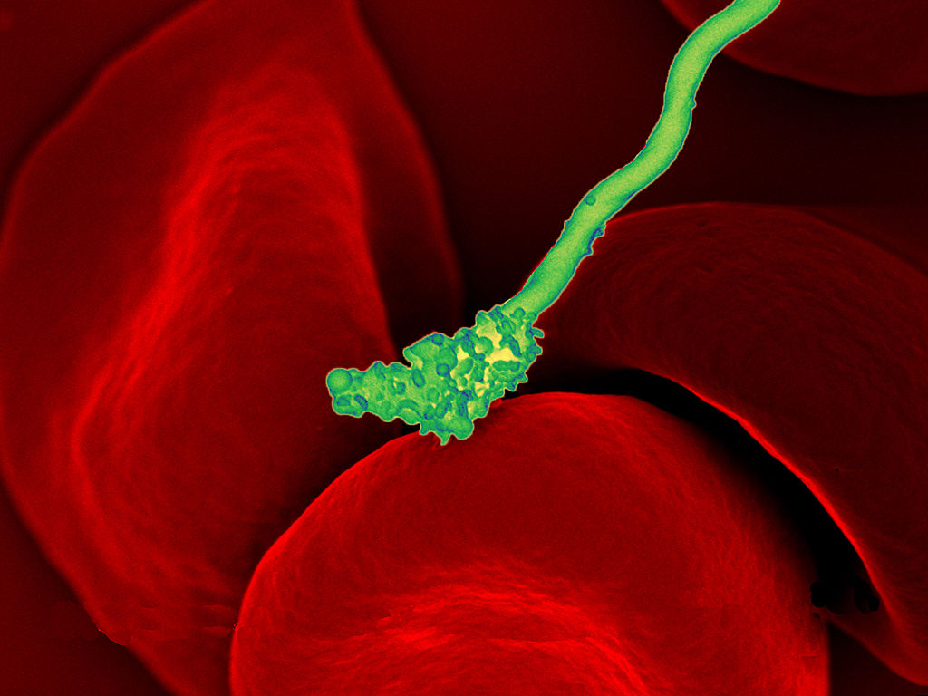
The colourful image in Figure 4.3.1 shows a bacterial cell (in green) attacking human red blood cells. The bacterium causes a disease called relapsing fever. The bacterial and human cells look very different in size and shape. Although all living cells have certain things in common — such as a plasma membrane and cytoplasm — different types of cells, even within the same organism, may have their own unique structures and functions. Cells with different functions generally have different shapes that suit them for their particular job. Cells vary not only in shape, but also in size, as this example shows. In most organisms, however, even the largest cells are no bigger than the period at the end of this sentence. Why are cells so small?
Explaining Cell Size
Most organisms, even very large ones, have microscopic cells. Why don’t cells get bigger instead of remaining tiny and multiplying? Why aren’t you one giant cell rolling around school? What limits cell size?
Once you know how a cell functions, the answers to these questions are clear. To carry out life processes, a cell must be able to quickly pass substances in and out of the cell. For example, it must be able to pass nutrients and oxygen into the cell and waste products out of the cell. Anything that enters or leaves a cell must cross its outer surface. The size of a cell is limited by its need to pass substances across that outer surface.
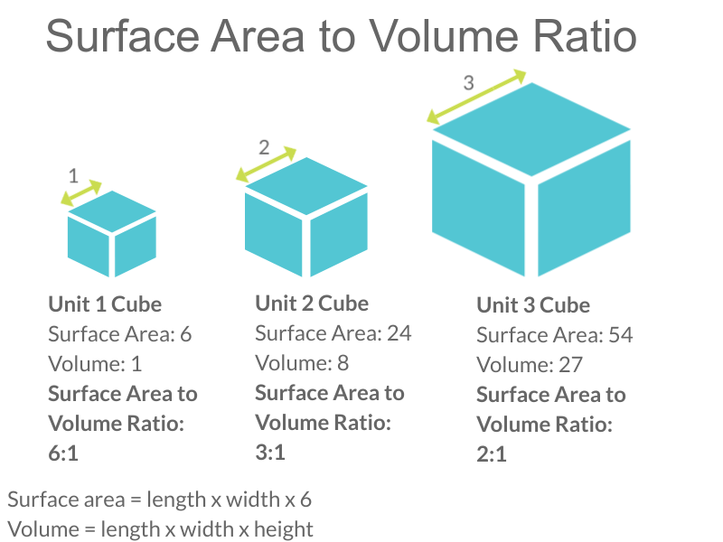
Look at the three cubes in Figure 4.3.2. A larger cube has less surface area relative to its volume than a smaller cube. This relationship also applies to cells — a larger cell has less surface area relative to its volume than a smaller cell. A cell with a larger volume also needs more nutrients and oxygen, and produces more waste. Because all of these substances must pass through the surface of the cell, a cell with a large volume will not have enough surface area to allow it to meet its needs. The larger the cell is, the smaller its ratio of surface area to volume, and the more difficult it will be for the cell to get rid of its waste and take in necessary substances. This is what limits the size of the cell.
Cell Form and Function
Cells with different functions often have varying shapes. The cells pictured below (Figure 4.3.3) are just a few examples of the many different shapes that human cells may have. Each type of cell has characteristics that help it do its job. The job of the nerve cell, for example, is to carry messages to other cells. The nerve cell has many long extensions that reach out in all directions, allowing it to pass messages to many other cells at once. Do you see the tail of each tiny sperm cell? Its tail helps a sperm cell “swim” through fluids in the female reproductive tract in order to reach an egg cell. The white blood cell has the job of destroying bacteria and other pathogens. It is a large cell that can engulf foreign invaders.
Figure 4.3.3 Human cells may have many different shapes that help them to do their jobs.
Cells With and Without a Nucleus
The nucleus is a basic cell structure present in many — but not all — living cells. The nucleus of a cell is a structure in the cytoplasm that is surrounded by a membrane (the nuclear membrane) and contains DNA. Based on whether or not they have a nucleus, there are two basic types of cells: prokaryotic cells and eukaryotic cells.
Prokaryotic Cells
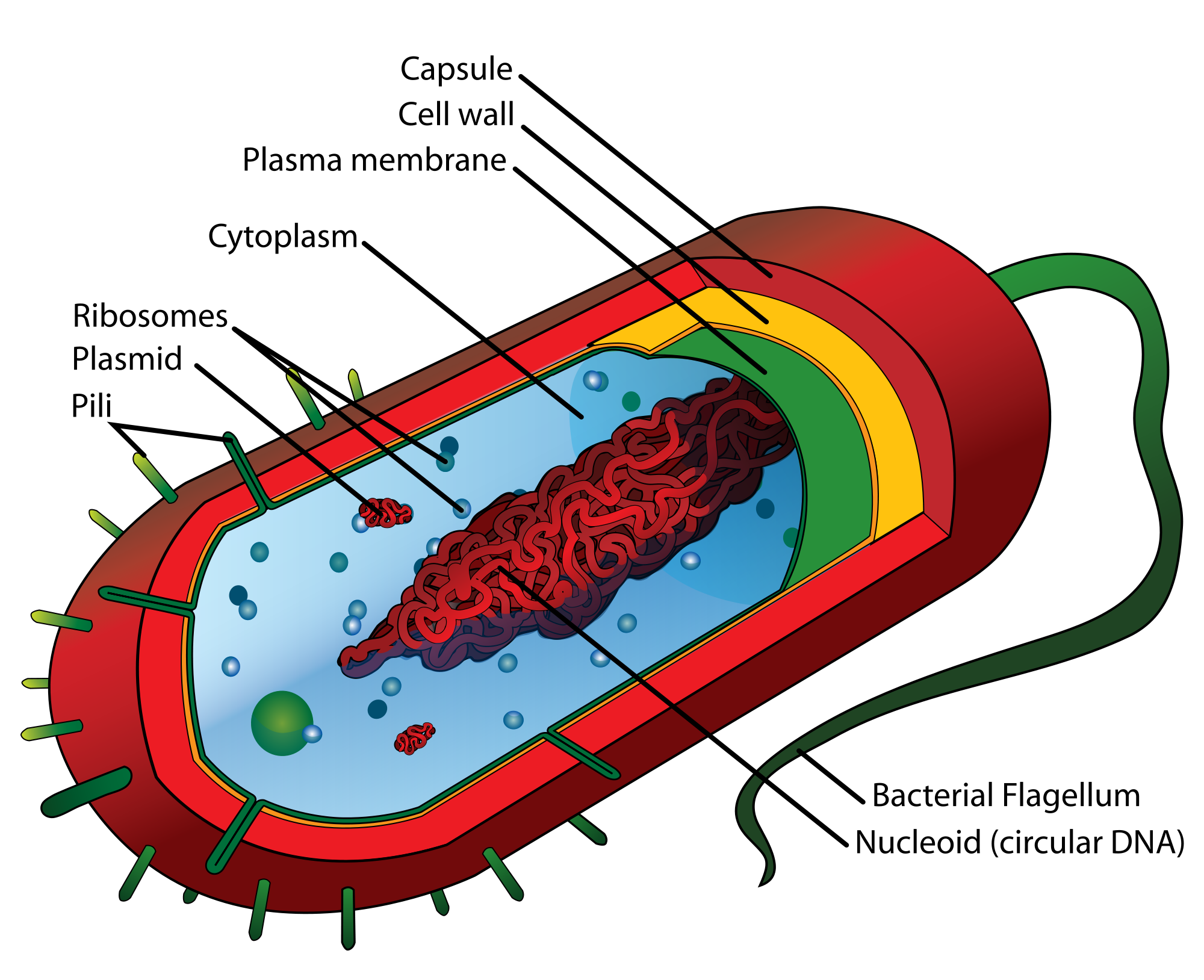
Prokaryotic cells are cells without a nucleus. The DNA in prokaryotic cells is in the cytoplasm, rather than enclosed within a nuclear membrane. In addition, these cells are typically smaller than eukaryotic cells and contain fewer organelles. Prokaryotic cells are found in single-celled organisms, such as the bacterium represented by the model in Figure 4.3.3. Organisms with prokaryotic cells are called prokaryotes. They were the first type of organisms to evolve, and they are still the most common organisms today.
Eukaryotic Cells
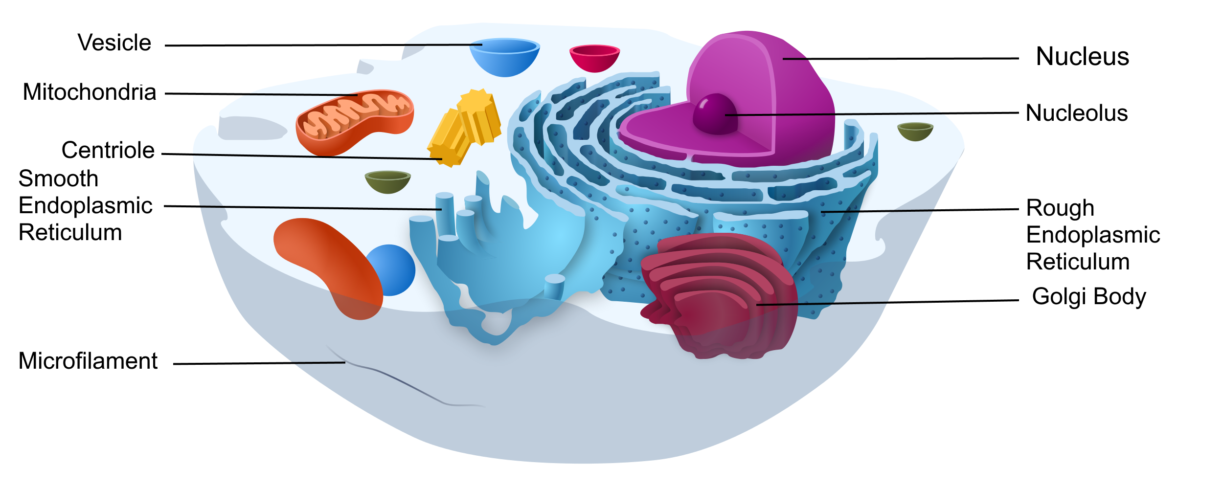
Eukaryotic cells are cells that contain a nucleus. A typical eukaryotic cell is represented by the model in Figure 4.3.4. Eukaryotic cells are usually larger than prokaryotic cells. They are found in some single-celled and all multicellular organisms. Organisms with eukaryotic cells are called eukaryotes, and they range from fungi to humans.
Besides a nucleus, eukaryotic cells also contain other organelles. An organelle is a structure within the cytoplasm that performs a specific job in the cell. Organelles called mitochondria, for example, provide energy to the cell, and organelles called vesicles store substances in the cell. Organelles allow eukaryotic cells to carry out more functions than prokaryotic cells can.
Interestingly, scientists think that mitochondria were once free-living prokaryotes that infected (or were engulfed by) larger cells. The two organisms developed a symbiotic relationship that was beneficial to both of them, resulting in the smaller prokaryote becoming an organelle within the larger cell. This is called endosymbiotic theory, and it is supported by a lot of evidence, including the fact that mitochondria have their own DNA separate from the DNA in the nucleus of the eukaryotic cell. Endosymbiotic theory will be described in more detail in later sections, and it’s also discussed in the video below.
Endosymbiotic Theory, Amoeba Sisters, 2017.
4.3 Summary
- Cells must be very small so they have a large enough surface area-to-volume ratio to maintain normal cell processes.
- Cells with different functions often have different shapes.
- Prokaryotic cells do not have a nucleus. Eukaryotic cells do have a nucleus, along with other organelles.
4.3 Review Questions
- Explain why most cells are very small.
- Discuss variations in the form and function of cells.
-
-
- Do human cells have organelles? Explain your answer.
- Which are usually larger – prokaryotic or eukaryotic cells? What do you think this means for their relative ability to take in needed substances and release wastes? Discuss your answer.
- DNA in eukaryotes is enclosed within the _______ ________.
- Name three different types of cells in humans.
- Which organelle provides energy in eukaryotic cells?
- What is a function of a vesicle in a cell?
4.3 Explore More
How we think complex cells evolved – Adam Jacobson, TED-Ed, 2015.
Prokaryotic vs. Eukaryotic Cells (updated), Amoeba Sisters, 2018.
Attributions
Figure 4.3.1
Borrelia_hermsii_Bacteria_(13758011613) by NAID on Wikimedia Commons is released into the public domain (https://en.wikipedia.org/wiki/Public_domain).
Figure 4.3.2
Cell Size by Christine Miller is released into the Public Domain (https://creativecommons.org/publicdomain/mark/1.0/).
Figure 4.3.3
- Chondrocyte. BioTek-Wikipedia-Image by BioTek Instruments, Inc. on Wikimedia Commons is used under a CC BY-SA 3.0 (https://creativecommons.org/licenses/by-sa/3.0/deed.en) license.
- Neutrophil with anthrax copy by Volker Brinkmann from PLOS Pathogens on Wikimedia Commons is used under a CC BY 2.5 (https://creativecommons.org/licenses/by/2.5/deed.en) license.
- PLoSBiol4.e126.Fig6fNeuron by Lee, et al. from PLOS Biology on Wikimedia Commons is used under a CC BY 2.5 (https://creativecommons.org/licenses/by/2.5/deed.en) license.
- Sperm (265 33) human by Doc. RNDr. Josef Reischig, CSc. on Wikimedia Commons is used under a CC BY-SA 3.0 (https://creativecommons.org/licenses/by-sa/3.0) license.
Figure 4.3.4
Model of a prokaryotic cell: bacterium by Mariana Ruiz Villarreal [LadyofHats] on Wikimedia Commons is released into the public domain (https://en.wikipedia.org/wiki/Public_domain).
Figure 4.3.5
Animal Cell adapted by Christine Miller is used under a CC0 1.0 (https://creativecommons.org/publicdomain/zero/1.0/deed.en) public domain dedication license. (Original image, Animal Cell Unannotated, is by Kelvin Song on Wikimedia Commons.)
References
Amoeba Sisters. (2017, May 3). Endosymbiotic theory. YouTube. https://www.youtube.com/watch?v=FGnS-Xk0ZqU&feature=youtu.be
Amoeba Sisters. (2018, July 30). Prokaryotic vs. eukaryotic cells (updated). YouTube. https://www.youtube.com/watch?v=Pxujitlv8wc&feature=youtu.be
Brinkmann, V. (November 2005). Neutrophil engulfing Bacillus anthracis. PLoS Pathogens 1 (3): Cover page [digital image]. DOI:10.1371. https://journals.plos.org/plospathogens/issue?id=10.1371/issue.ppat.v01.i03
Lee, W.C.A., Huang, H., Feng, G., Sanes, J.R., Brown, E.N., et al. (2005, December 27) Figure 6f, slightly altered (plus scalebar, minus letter “f”.) [digital image]. Dynamic Remodeling of Dendritic Arbors in GABAergic Interneurons of Adult Visual Cortex. PLoS Biology, 4(2), e29. doi:10.1371/journal.pbio.0040029. https://journals.plos.org/plosbiology/article?id=10.1371/journal.pbio.0040029
TED-Ed. (2015, February 17). How we think complex cells evolved – Adam Jacobson. https://www.youtube.com/watch?v=9i7kAt97XYU&feature=youtu.be
A central organelle containing hereditary material.
Deoxyribonucleic acid - the molecule carrying genetic instructions for the development, functioning, growth and reproduction of all known organisms and many viruses.
Cells which lack membrane-bound structures, specifically a nucleus. Instead they generally have a single circular chromosome located in an area of the cell called the nucleoid.
Cells which have a nucleus enclosed within membranes, unlike prokaryotes, which have no membrane-bound organelles.
A tiny cellular structure that performs specific functions within a cell.
A double-membrane-bound organelle found in most eukaryotic organisms. Mitochondria convert oxygen and nutrients into adenosine triphosphate (ATP). ATP is the chemical energy "currency" of the cell that powers the cell's metabolic activities.
The ability to do work.

.jpg)
_human.jpg)