8.3 Neurons and Neuroglia
Created by CK-12 Foundation/Adapted by Christine Miller
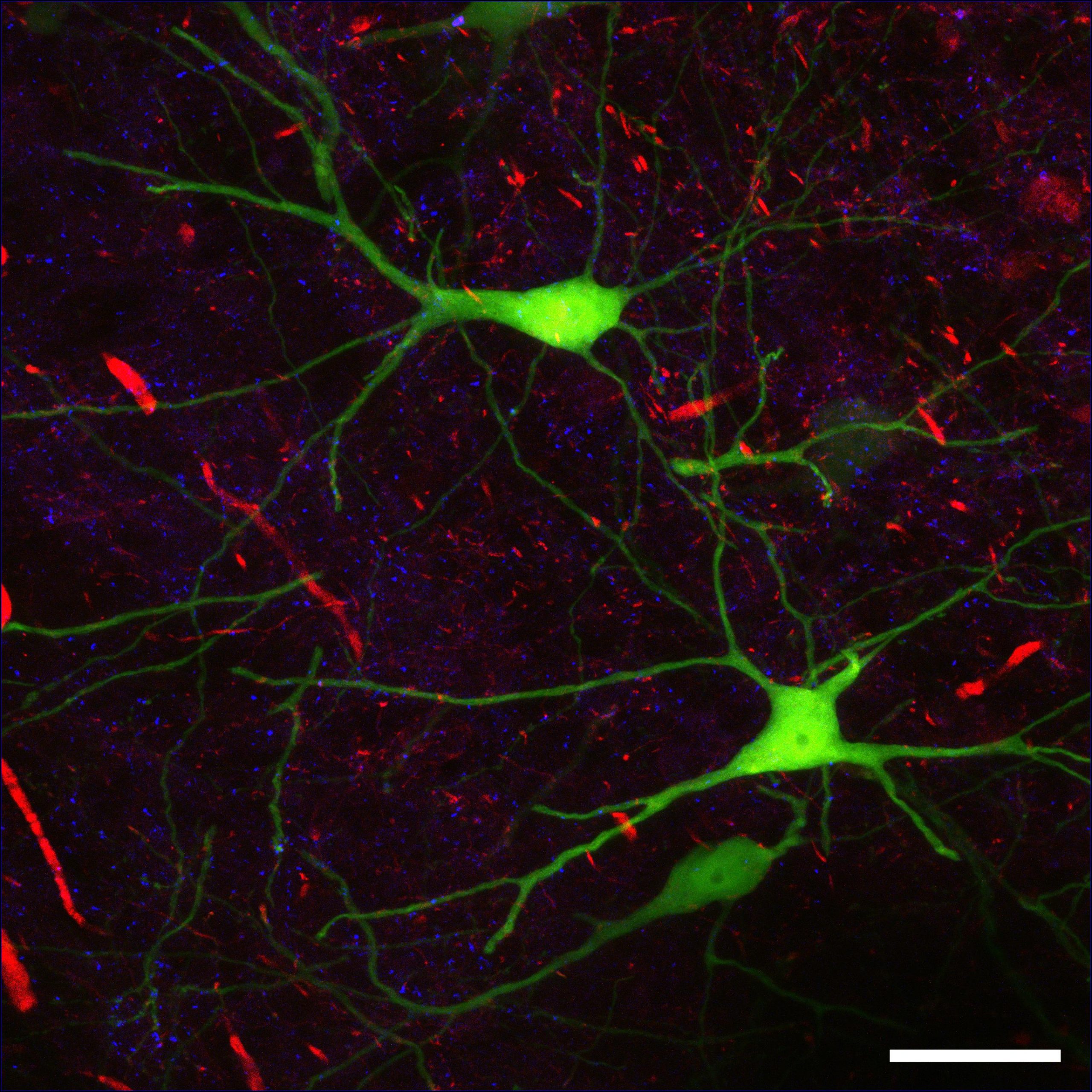
Life as Art
This colourful picture (Figure 8.3.1) could be an abstract work of modern art. You might imagine it hanging in an art museum or art gallery. In fact, the picture illustrates real life — not artistic creation. It is a micrograph of human nervous tissue. The neon green structures in the picture are neurons. The neuron is one of two basic types of cells in the nervous system. The other type is the neuroglial cell.
Neurons
Neurons — also called nerve cells — are electrically excitable cells that are the main functional units of the nervous system. Their function is to transmit nerve impulses, and they are the only type of human cells that can carry out this function.
Neuron Structure
Figure 8.3.2 shows the structure of a typical neuron. Click on each of the main parts to learn about their functions.
Figure 8.3.2 The structure of a typical neuron.
Neurogenesis
Fully differentiated neurons, with all their special structures, cannot divide and form new daughter neurons. Until recently, scientists thought that new neurons could no longer be formed after the brain developed prenatally. In other words, they thought that people were born with all the brain neurons they would ever have, and as neurons died, they would not be replaced. However, new evidence shows that additional neurons can form in the brain, even in adults, from the division of undifferentiated neural stem cells found throughout the brain. The production of new neurons is called neurogenesis. The extent to which it can occur is not known, but it is not likely to be very great in humans.
Neurons in Nervous Tissues
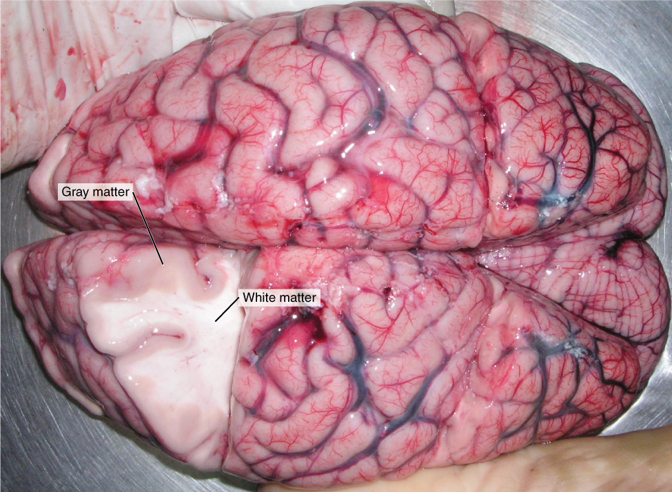
The nervous tissue in the brain and spinal cord consists of gray matter and white matter. Gray matter contains mainly non-myelinated structures, including the cell bodies and dendrites of neurons. It is gray only in cadavers. Living gray matter is actually more pink than gray (see Figure 8.3.3) White matter consists mainly of axons covered with a myelin sheath, which gives them their white colour. White matter also makes up the nerves of the peripheral nervous system. Nerves consist of long bundles of myelinated axons that extend to muscles, organs, or glands throughout the body. The axons in each nerve are bundled together like wires in a cable. Axons in nerves may be more than a metre long in an adult. The longest nerve runs from the base of the spine to the toes.
Types of Neurons
There are hundreds of different types of neurons in the human nervous system that exhibit a variety of structures and functions. Nonetheless, many neurons can be classified functionally based on the direction in which they carry nerve impulses.
- Sensory (also called afferent) neurons carry nerve impulses from sensory receptors in tissues and organs to the central nervous system. They change physical stimuli (such as touch, light, and sound) into nerve impulses.
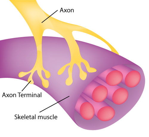
- Motor (also called efferent) neurons, like the one in the diagram below (Figure 8.3.4), carry nerve impulses from the central nervous system to muscles and glands. They change nerve signals into the activation of these structures.
- Within the spinal cord or brain, interneurons carry nerve impulses back and forth, often between sensory and motor neurons.
Neuroglia
In addition to neurons, nervous tissues also consist of neuroglia, also called glial cells. The root of the word glial comes from a Greek word meaning “glue,” which reflects earlier ideas about the role of neuroglia in nervous tissues. Neuroglia were thought to be little more than “glue” holding together the all-important neurons, but this is no longer the case. They are now known to play many vital roles in the nervous system. There are several different types of neuroglia, each with a different function. You can see six types of neuroglia in Figure 8.3.5.
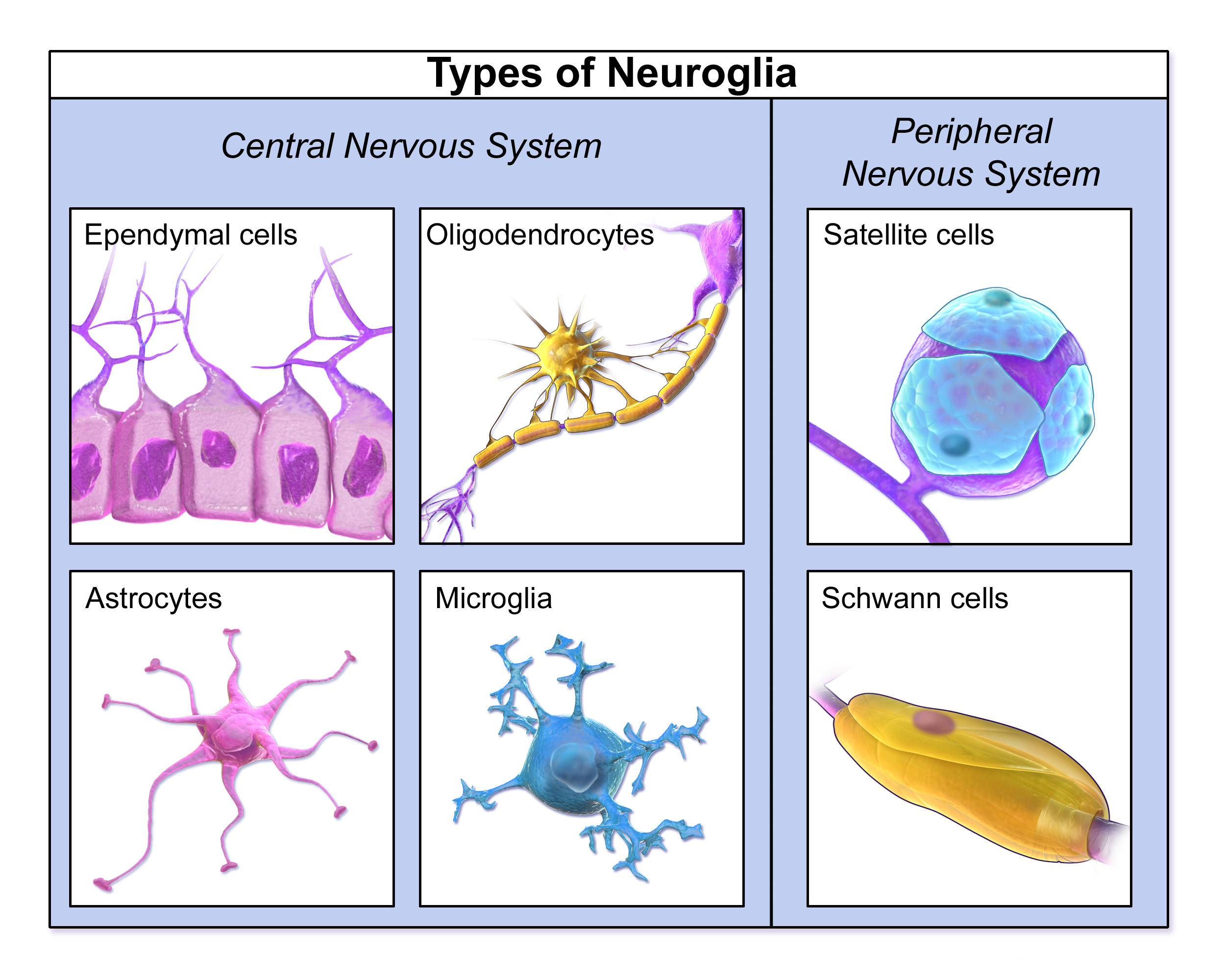
In general, neuroglia provide support for neurons and help them carry out the basic function of nervous tissues, which is to transmit nerve impulses. For example, oligodendrocytes in the central nervous system and Schwann cells in the peripheral nervous system generate the lipids that make up myelin sheaths, which increase the speed of nerve impulses’ transmission. Functions of other neuroglia cells include holding neurons in place, supplying neurons with nutrients, regulating the repair of neurons, destroying pathogens, removing dead neurons, and directing axons to their targets. Neuroglia may also play a role in the transmission of nerve impulses, but this is still under study. Unlike mature neurons, mature glial cells retain the ability to divide by undergoing mitosis.
In the human brain, there are generally roughly equal numbers of neurons and neuroglia. If you think intelligence depends on how many neurons you have, think again. Having a relatively high number of neuroglia is actually associated with higher intelligence. When Einstein’s brain was analyzed, researchers discovered a significantly higher-than-normal ratio of neuroglia to neurons in areas of the brain associated with mathematical processing and language. On an evolutionary scale, as well, an increase in the ratio of neuroglia to neurons is associated with greater intelligence in species.
Feature: My Human Body
Would you like your brain to make new neurons that could help you become a better learner? When it comes to learning new things, what college student wouldn’t want a little more brain power? If research about rats applies to humans, then sustained aerobic exercise (such as running) can increase neurogenesis in the adult brain, and specifically in the hippocampus, a brain structure important for learning temporally and/or spatially complex tasks, as well as memory. Although the research is still at the beginning stages, it suggests that exercise may actually lead to a “smarter” brain. Even if the research results are not ultimately confirmed for humans, though, it can’t hurt to get more aerobic exercise. It is certainly beneficial for your body, if not your brain!
8.3 Summary
- Neurons are one of two major types of nervous system cells. They are electrically excitable cells that transmit nerve impulses.
- Neuroglia are the other major type of nervous system cells. There are many types of neuroglia and they have many specific functions. In general, neuroglia function to support, protect, and nourish neurons.
- The main parts of a neuron include the cell body, dendrites, and axon. The cell body contains the nucleus. Dendrites receive nerve impulses from other cells, and the axon transmits nerve impulses to other cells at axon terminals. A synapse is a complex membrane junction at the end of an axon terminal that transmits signals to another cell.
- Axons are often wrapped in an electrically-insulating myelin sheath, which is produced by neuroglia. Electrical signals occur at gaps in the myelin sheath, called nodes of Ranvier, which speeds the conduction of nerve impulses down the axon.
- Neurogenesis, or the formation of new neurons by cell division, may occur in a mature human brain, but only to a limited extent.
- The nervous tissue in the brain and spinal cord consists of gray matter (which contains unmyelinated cell bodies and dendrites of neurons) and white matter (which contains mainly myelinated axons of neurons). Nerves of the peripheral nervous system consist of long bundles of myelinated axons that extend throughout the body.
- There are hundreds of types of neurons in the human nervous system, but many can be classified on the basis of the direction in which they carry nerve impulses. Sensory neurons carry nerve impulses away from the body and toward the central nervous system, motor neurons carry them away from the central nervous system and toward the body, and interneurons often carry them between sensory and motor neurons.
8.3 Review Questions
-
- Describe the myelin sheath and nodes of Ranvier. How does their arrangement allow nerve impulses to travel very rapidly along axons?
- Define neurogenesis. What is the potential for neurogenesis in the human brain?
- Relate neurons to different types of nervous tissues.
- Compare and contrast sensory and motor neurons.
- Identify the role of interneurons.
- Identify four specific functions of neuroglia.
- What is the relationship between the proportion of neuroglia to neurons and intelligence?
-
-
8.3 Explore More
Thriving in the Face of Adversity | Stephanie Buxhoeveden | TEDxHerndon, TEDx Talks, 2015.
You can grow new brain cells. Here’s how | Sandrine Thuret,
TED, 2015.
Attributions
Figure 8.3.1
Nervous Tissue Confocal Microscopy/ Mouse brain, confocal microscopy by ZEISS Microscopy on Flickr is used under a CC BY-NC-ND 2.0 (https://creativecommons.org/licenses/by-nc-nd/2.0/) license.
Figure 8.3.2
Parts of a Neuron by Open Stax on Wikimedia Commons is used and adapted by Christine Miller under the CC BY 4.0 (https://creativecommons.org/licenses/by/4.0/) license.
Figure 8.3.3
White_and_Gray_Matter by OpenStax on Wikimedia Commons is used under a CC BY 4.0 (https://creativecommons.org/licenses/by/4.0/) license.
Figure 8.3.4
Neuromuscular Junction by CK-12 Foundation is used under a CC BY-NC 3.0 (https://creativecommons.org/licenses/by-nc/3.0/) license.
Figure 8.3.5
TypesofNeuroglia by BruceBlaus on Wikimedia Commons is used under a CC BY 3.0 (https://creativecommons.org/licenses/by/3.0/deed.en) license.
References
Betts, J. G., Young, K.A., Wise, J.A., Johnson, E., Poe, B., Kruse, D.H., Korol, O., Johnson, J.E., Womble, M., DeSaix, P. (2016, May 18). Figure 12.3 Gray matter and white matter [digital image]. In Anatomy and Physiology (Section 12.1). OpenStax. https://openstax.org/books/anatomy-and-physiology/pages/12-1-basic-structure-and-function-of-the-nervous-system
Betts, J. G., Young, K.A., Wise, J.A., Johnson, E., Poe, B., Kruse, D.H., Korol, O., Johnson, J.E., Womble, M., DeSaix, P. (2016, May 18). Figure 12.8 Parts of a neuron [digital image]. In Anatomy and Physiology (Section 12.2). OpenStax. https://openstax.org/books/anatomy-and-physiology/pages/12-2-nervous-tissue
Blausen.com staff. (2014). Types of neuroglia cells [digital image]. Medical gallery of Blausen Medical 2014. WikiJournal of Medicine, 1 (2). DOI:10.15347/wjm/2014.010. ISSN 2002-4436. Wikiversity.org. https://en.wikiversity.org/wiki/WikiJournal_of_Medicine/Medical_gallery_of_Blausen_Medical_2014
Brainard, J/ CK-12 Foundation. (2016). Figure 3 The axon in this diagram is part of a motor neuron. [digital image]. In CK-12 College Human Biology (Section 10.3) [online Flexbook]. CK12.org. https://www.ck12.org/book/ck-12-college-human-biology/section/10.3/
TED. (2015, October 30). You can grow new brain cells. Here’s how | Sandrine Thuret. YouTube. https://www.youtube.com/watch?v=B_tjKYvEziI&feature=youtu.be
TEDx Talks. (2015, April 3). Thriving in the face of adversity | Stephanie Buxhoeveden | TEDxHerndon. YouTube. https://www.youtube.com/watch?v=zuLOT6GsAxw&feature=youtu.be
A specialized tissue found in the central nervous system and the peripheral nervous system. It consists of neurons and supporting cells called neuroglia. The nervous system is responsible for the control of the body and the communication among its parts.
A functional unit of the nervous system that transmits nerve impulses; also called a nerve cell.
A class of nervous system cell that provides support for neurons and helps them transmit nerve impulses.
The highly complex body system of an animal that coordinates its actions and sensory information by transmitting signals to and from different parts of its body. The nervous system detects environmental changes that impact the body, then works in tandem with the endocrine system to respond to such events.
A signal transmitted along a nerve fiber.
The formation of new neurons by cell division.
The central nervous system organ inside the skull that is the control center of the nervous system.
A thin, tubular bundle of central nervous system tissue that extends from the brainstem down the back to the pelvis and connects the brain with the peripheral nervous system.
A type of nervous tissue that is found only in the brain and spinal cord and consists mainly of un-myelinated cell bodies and dendrites of neurons.
A type of nervous tissue that consists mainly of the myelinated axons of neurons.
The lipid layer around the axon of a neuron that allows nerve impulses to travel more rapidly down the axon.
A structure in the nervous system that consists of cable-like bundles of axons and makes up the majority of the peripheral nervous system.
One of two major divisions of the nervous system that consists of all the nervous tissue that lies outside the central nervous system.
A long extension of the cell body of a neuron that transmits nerve impulses to other cells.
Type of neuron that carries nerve impulses from sensory receptors in tissues and organs to the central nervous system; also called afferent neuron.
Specialized nerve cell that responds to a particular type of stimulus such as light or chemicals by generating a nerve impulse.
A cellular organizational level between cells and a complete organ. A tissue is an ensemble of similar cells and their extracellular matrix from the same origin that together carry out a specific function. Organs are then formed by the functional grouping together of multiple tissues.
A group of tissues in a living organism that have been adapted to perform a specific function. In higher animals, organs are grouped into organ systems; e.g., the esophagus, stomach, and liver are organs of the digestive system.
One of two main divisions of the nervous system that includes the brain and spinal cord.
A type of neuron that carries nerve impulses from the central nervous system to muscles and glands; also called efferent neuron.
A type of neuron that carries nerve impulses between other neurons, often between sensory and motor neurons.
A nervous system cell that provides support for neurons and helps them transmit nerve impulses.
A type of neuroglia whose main functions are to provide support and insulation to axons in the central nervous system of some vertebrates, equivalent to the function performed by Schwann cells in the peripheral nervous system.
A variety of neuroglia that keep peripheral nerve fibres (both myelinated and unmyelinated) alive. In myelinated axons, Schwann cells form the myelin sheath.
The central part of a neuron that contains the nucleus and other cell organelles.
An extension of the cell body of a neuron that receives nerve impulses from other neurons. A neuron will have several dendrites extending from the cell body.
A central organelle containing hereditary material.
The place where the axon terminal of a neuron transmits a chemical or electrical signal to another cell.
One of the regularly spaced gaps in the myelin sheath along an axon that allows the action potential (electrical signal) to travel very rapidly.
