15.6 Accessory Organs of Digestion
Created by CK-12 Foundation/Adapted by Christine Miller

Jaundiced Eyes
Did you ever hear of a person looking at something or someone with a “jaundiced eye”? It means to take a negative view, such as envy, maliciousness, or ill will. The expression may be based on the antiquated idea that liver bile is associated with such negative emotions as these, as well as the fact that excessive liver bile causes jaundice, or yellowing of the eyes and skin. Jaundice is likely a sign of a liver disorder or blockage of the duct that carries bile away from the liver. Bile contains waste products, making the liver an organ of excretion. Bile has an important role in digestion, which makes the liver an accessory organ of digestion, too.
What Are Accessory Organs of Digestion?
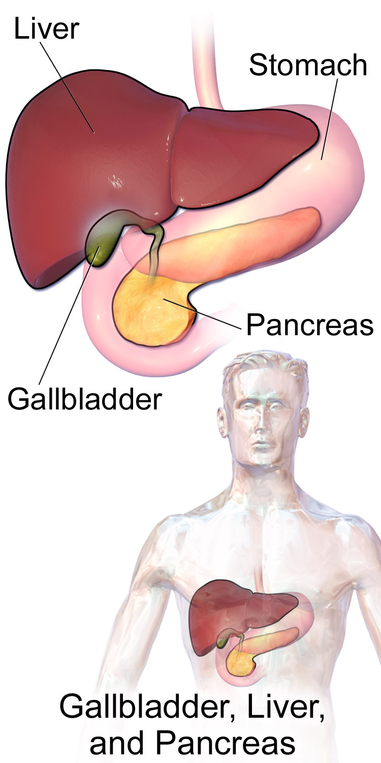
Accessory organs of digestion are organs that secrete substances needed for the chemical digestion of food, but through which food does not actually pass as it is digested. Besides the liver, the major accessory organs of digestion are the gallbladder and pancreas. These organs secrete or store substances that are needed for digestion in the first part of the small intestine — the duodenum — where most chemical digestion takes place. You can see the three organs and their locations in Figure 15.6.2.
Liver
The liver is a vital organ located in the upper right part of the abdomen. It lies just below the diaphragm, to the right of the stomach. The liver plays an important role in digestion by secreting bile, but the liver has a wide range of additional functions unrelated to digestion. In fact, some estimates put the number of functions of the liver at about 500! A few of them are described below.
Structure of the Liver
The liver is a reddish brown, wedge-shaped structure. In adults, the liver normally weighs about 1.5 kg (about 3.3 lb). It is both the heaviest internal organ and the largest gland in the human body. The liver is divided into four lobes of unequal size and shape. Each lobe, in turn, is made up of lobules, which are the functional units of the liver. Each lobule consists of millions of liver cells, called hepatic cells (or hepatocytes). They are the basic metabolic cells that carry out the various functions of the liver.
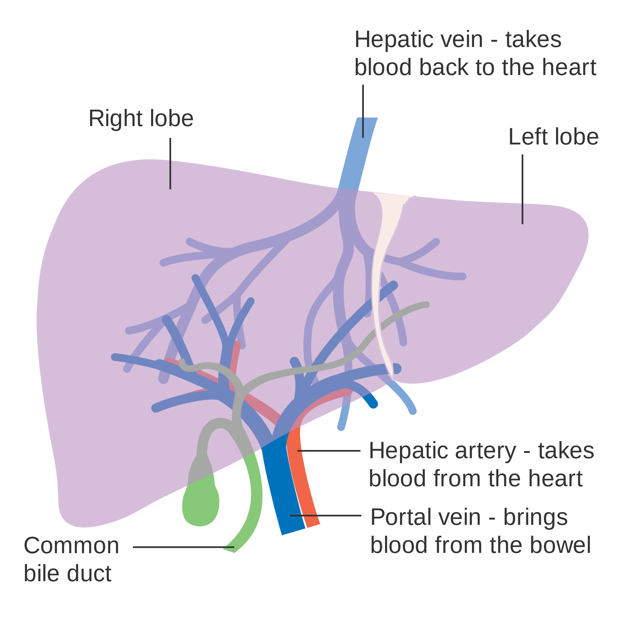
As shown in Figure 15.6.3, the liver is connected to two large blood vessels: the hepatic artery and the portal vein. The hepatic artery carries oxygen-rich blood from the aorta, whereas the portal vein carries blood that is rich in digested nutrients from the GI tract and wastes filtered from the blood by the spleen. The blood vessels subdivide into smaller arteries and capillaries, which lead into the liver lobules. The nutrients from the GI tract are used to build many vital biochemical compounds, and the wastes from the spleen are degraded and excreted.
Functions of the Liver
The main digestive function of the liver is the production of bile. Bile is a yellowish alkaline liquid that consists of water, electrolytes, bile salts, and cholesterol, among other substances, many of which are waste products. Some of the components of bile are synthesized by hepatocytes. The rest are extracted from the blood.
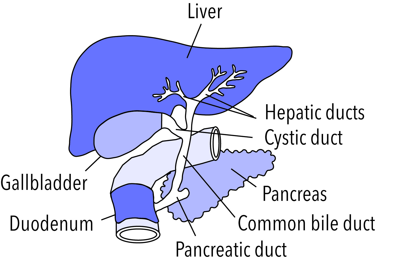
As shown in Figure 15.6.4, bile is secreted into small ducts that join together to form larger ducts, with just one large duct carrying bile out of the liver. If bile is needed to digest a meal, it goes directly to the duodenum through the common bile duct. In the duodenum, the bile neutralizes acidic chyme from the stomach and emulsifies fat globules into smaller particles (called micelles) that are easier to digest chemically by the enzyme lipase. Bile also aids with the absorption of vitamin K. Bile that is secreted when digestion is not taking place goes to the gallbladder for storage until the next meal. In either case, the bile enters the duodenum through the common bile duct.
Besides its roles in digestion, the liver has many other vital functions:
- The liver synthesizes glycogen from glucose and stores the glycogen as required to help regulate blood sugar levels. It also breaks down the stored glycogen to glucose and releases it back into the blood as needed.
- The liver stores many substances in addition to glycogen, including vitamins A, D, B12, and K. It also stores the minerals iron and copper.
- The liver synthesizes numerous proteins and many of the amino acids needed to make them. These proteins have a wide range of functions. They include fibrinogen, which is needed for blood clotting; insulin-like growth factor (IGF-1), which is important for childhood growth; and albumen, which is the most abundant protein in blood serum and functions to transport fatty acids and steroid hormones in the blood.
- The liver synthesizes many important lipids, including cholesterol, triglycerides, and lipoproteins.
- The liver is responsible for the breakdown of many waste products and toxic substances. The wastes are excreted in bile or travel to the kidneys, which excrete them in urine.
The liver is clearly a vital organ that supports almost every other organ in the body. Because of its strategic location and diversity of functions, the liver is also prone to many diseases, some of which cause loss of liver function. There is currently no way to compensate for the absence of liver function in the long term, although liver dialysis techniques can be used in the short term. An artificial liver has not yet been developed, so liver transplantation may be the only option for people with liver failure.
Gallbladder
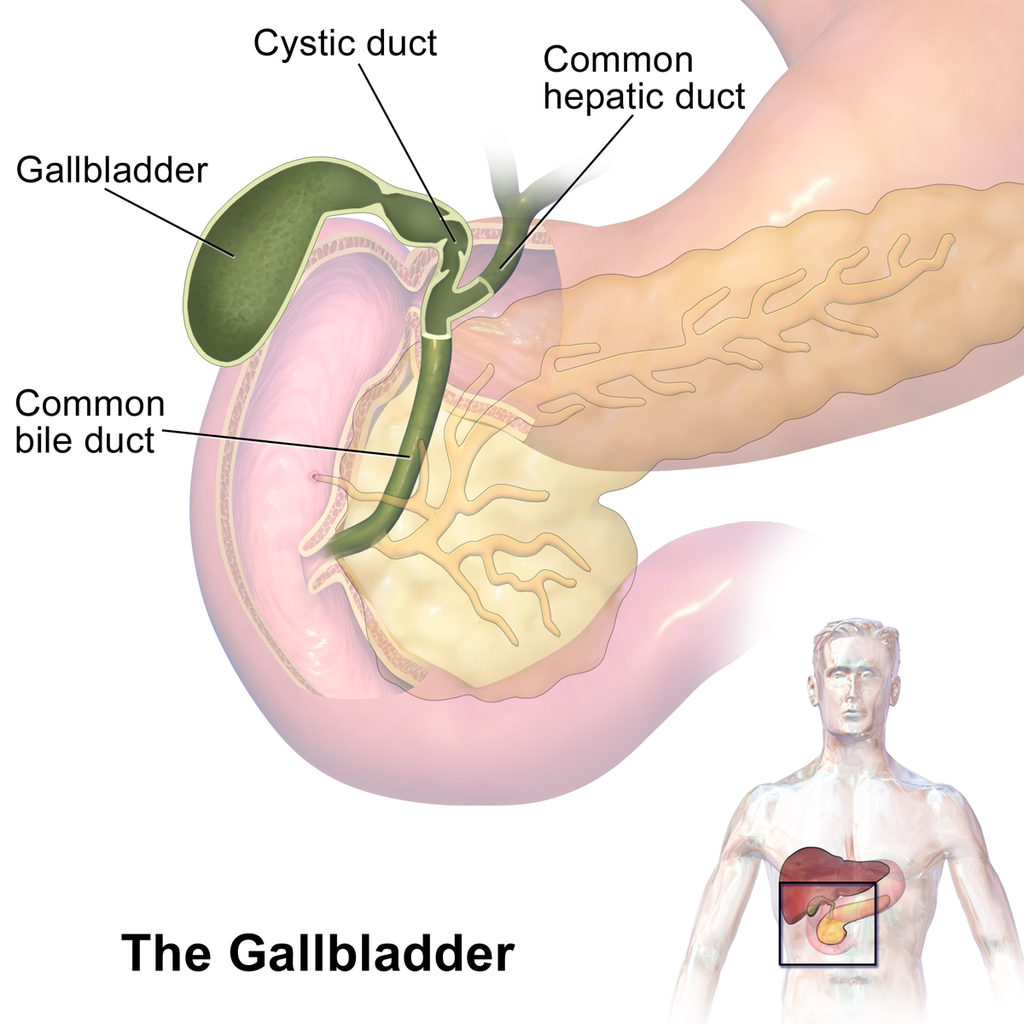
The gallbladder is a small, hollow, pouch-like organ that lies just under the right side of the liver (see Figure 15.6.5). It is about 8 cm (about 3 in) long and shaped like a tapered sac, with the open end continuous with the cystic duct. The gallbladder stores and concentrates bile from the liver until it is needed in the duodenum to help digest lipids. After the bile leaves the liver, it reaches the gallbladder through the cystic duct. At any given time, the gallbladder may store between 30 to 60 mL (1 to 2 oz) of bile. A hormone stimulated by the presence of fat in the duodenum signals the gallbladder to contract and force its contents back through the cystic duct and into the common bile duct to drain into the duodenum.
Pancreas
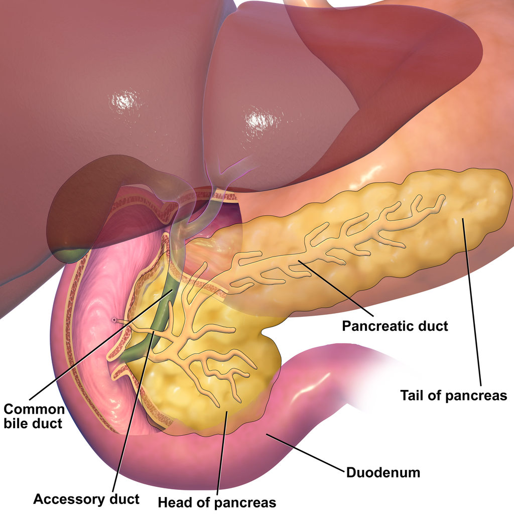
The pancreas is a glandular organ that is part of both the digestive system and the endocrine system. As shown in Figure 15.6.6, it is located in the abdomen behind the stomach, with the head of the pancreas surrounded by the duodenum of the small intestine. The pancreas is about 15 cm (almost 6 in) long, and it has two major ducts: the main pancreatic duct and the accessory pancreatic duct. Both of these ducts drain into the duodenum.
As an endocrine gland, the pancreas secretes several hormones, including insulin and glucagon, which circulate in the blood. The endocrine hormones are secreted by clusters of cells called pancreatic islets (or islets of Langerhans). As a digestive organ, the pancreas secretes many digestive enzymes and also bicarbonate, which helps neutralize acidic chyme after it enters the duodenum. The pancreas is stimulated to secrete its digestive substances when food in the stomach and duodenum triggers the release of endocrine hormones into the blood that reach the pancreas via the bloodstream. The pancreatic digestive enzymes are secreted by clusters of cells called acini, and they travel through the pancreatic ducts to the duodenum. In the duodenum, they help to chemically break down carbohydrates, proteins, lipids, and nucleic acids in chyme. The pancreatic digestive enzymes include:
- Amylase, which helps digest starch and other carbohydrates.
- Trypsin and chymotrypsin, which help digest proteins.
- Lipase, which helps digest lipids.
- Deoxyribonucleases and ribonucleases, which help digest nucleic acids.
15.6 Summary
- Accessory organs of digestion are organs that secrete substances needed for the chemical digestion of food, but through which food does not actually pass as it is digested. The accessory organs include the liver, gallbladder, and pancreas. These organs secrete or store substances that are carried to the duodenum of the small intestine as needed for digestion.
- The liver is a large organ in the abdomen that is divided into lobes and smaller lobules, which consist of metabolic cells called hepatic cells, or hepatocytes. The liver receives oxygen in blood from the aorta through the hepatic artery. It receives nutrients in blood from the GI tract and wastes in blood from the spleen through the portal vein.
- The main digestive function of the liver is the production of the alkaline liquid called bile. Bile is carried directly to the duodenum by the common bile duct or to the gallbladder first for storage. Bile neutralizes acidic chyme that enters the duodenum from the stomach, and also emulsifies fat globules into smaller particles (micelles) that are easier to digest chemically.
- Other vital functions of the liver include regulating blood sugar levels by storing excess sugar as glycogen, storing many vitamins and minerals, synthesizing numerous proteins and lipids, and breaking down waste products and toxic substances.
- The gallbladder is a small pouch-like organ near the liver. It stores and concentrates bile from the liver until it is needed in the duodenum to neutralize chyme and help digest lipids.
- The pancreas is a glandular organ that secretes both endocrine hormones and digestive enzymes. As an endocrine gland, the pancreas secretes insulin and glucagon to regulate blood sugar. As a digestive organ, the pancreas secretes digestive enzymes into the duodenum through ducts. Pancreatic digestive enzymes include amylase (starches) trypsin and chymotrypsin (proteins), lipase (lipids), and ribonucleases and deoxyribonucleases (RNA and DNA).
15.6 Review Questions
- Name three accessory organs of digestion. How do these organs differ from digestive organs that are part of the GI tract?
-
- Describe the liver and its blood supply.
- Explain the main digestive function of the liver and describe the components of bile and it’s importance in the digestive process.
- What type of secretions does the pancreas release as part of each body system?
- List pancreatic enzymes that work in the duodenum, along with the substances they help digest.
- What are two substances produced by accessory organs of digestion that help neutralize chyme in the small intestine? Where are they produced?
- People who have their gallbladder removed sometimes have digestive problems after eating high-fat meals. Why do you think this happens?
- Which accessory organ of digestion synthesizes cholesterol?
15.6 Explore More
What does the pancreas do? – Emma Bryce, TED-Ed. 2015.
What does the liver do? – Emma Bryce, TED-Ed, 2014.
Scar wars: Repairing the liver, nature video, 2018.
Attributions
Figure 15.6.1
Scleral_Icterus by Sheila J. Toro on Wikimedia Commons is used under a CC BY 4.0 (https://creativecommons.org/licenses/by/4.0) license.
Figure 15.6.2
Blausen_0428_Gallbladder-Liver-Pancreas_Location by BruceBlaus on Wikimedia Commons is used under a CC BY 3.0 (https://creativecommons.org/licenses/by/3.0) license.
Figure 15.6.3
Diagram_showing_the_two_lobes_of_the_liver_and_its_blood_supply_CRUK_376.svg by Cancer Research UK on Wikimedia Commons is used under a CC BY-SA 4.0 (https://creativecommons.org/licenses/by-sa/4.0) license.
Figure 15.6.4
Gallbladder by NIH Image Gallery on Flickr is used CC BY-NC 2.0 (https://creativecommons.org/licenses/by-nc/2.0/) license.
Figure 15.6.5
Gallbladder_(organ) (1) by BruceBlaus on Wikimedia Commons is used under a CC BY-SA 4.0 (https://creativecommons.org/licenses/by-sa/4.0) license. (See a full animation of this medical topic at blausen.com.)
Figure 15.6.6
Blausen_0698_PancreasAnatomy by BruceBlaus on Wikimedia Commons is used under a CC BY 3.0 (https://creativecommons.org/licenses/by/3.0) license.
References
Blausen.com Staff. (2014). Medical gallery of Blausen Medical 2014. WikiJournal of Medicine 1 (2). DOI:10.15347/wjm/2014.010. ISSN 2002-4436.
nature video. (2018, December 19). Scar wars: Repairing the liver. YouTube. https://www.youtube.com/watch?v=a0d1yvGcfzQ&feature=youtu.be
TED-Ed. (2014, November 25). What does the liver do? – Emma Bryce. YouTube. https://www.youtube.com/watch?v=wbh3SjzydnQ&feature=youtu.be
TED-Ed. (2015, February 19). What does the pancreas do? – Emma Bryce. YouTube. https://www.youtube.com/watch?v=8dgoeYPoE-0&feature=youtu.be
An organ of digestion and excretion that secretes bile for lipid digestion and breaks down excess amino acids and toxins in the blood.
A sac-like organ that stores bile from the liver and secretes it into the duodenum of the small intestine as needed for digestion.
A long, flat gland that sits tucked behind the stomach in the upper abdomen. The pancreas produces enzymes that help digestion and hormones that help regulate the way your body processes sugar (glucose).
The first and shortest of three parts of the small intestine where most chemical digestion occurs.
A large, dome-shaped muscle below the lungs that allows breathing to occur when it alternately contracts and relaxes.
A sac-like organ of the digestive system between the esophagus and small intestine in which both mechanical and chemical digestion take place.
Fluid produced by the liver and stored in the gall bladder that is secreted into the small intestine to help digest lipids and neutralize acid from the stomach.
A liver cell.
A multi-branched polysaccharide of glucose that serves as a form of energy storage in animals, fungi, and bacteria.
Glucose (also called dextrose) is a simple sugar with the molecular formula C6H12O6. Glucose is the most abundant monosaccharide, a subcategory of carbohydrates. Glucose is mainly made by plants and most algae during photosynthesis from water and carbon dioxide, using energy from sunlight.
A class of biological molecule consisting of linked monomers of amino acids and which are the most versatile macromolecules in living systems and serve crucial functions in essentially all biological processes.
Amino acids are organic compounds that combine to form proteins.
A lipid. Cholesterol and its derivatives are important constituents of cell membranes and precursors of other steroid compounds, but a high proportion in the blood of low-density lipoprotein (which transports cholesterol to the tissues) is associated with an increased risk of coronary heart disease.
A body system including a series of hollow organs joined in a long, twisting tube from the mouth to the anus. The hollow organs that make up the GI tract are the mouth, esophagus, stomach, small intestine, large intestine, and anus. The liver, pancreas, and gallbladder are the solid organs of the digestive system.
The body system which acts as a chemical messenger system comprising feedback loops of the hormones released by internal glands of an organism directly into the circulatory system, regulating distant target organs. In humans, the major endocrine glands are the thyroid gland and the adrenal glands.
A hormone is a signaling molecule produced by glands in multicellular organisms that target distant organs to regulate physiology and behavior.
A hormone made by the pancreas that allows your body to use sugar (glucose) from carbohydrates in the food that you eat for energy or to store glucose for future use.
A peptide hormone, produced by alpha cells of the pancreas. It works to raise the concentration of glucose and fatty acids in the bloodstream, and is considered to be the main catabolic hormone of the body. It is also used as a medication to treat a number of health conditions.
A thick, semi-liquid mixture that food in the gastrointestinal tract becomes by the time it leaves the stomach.
An enzyme, found chiefly in saliva and pancreatic fluid, that converts starch and glycogen into simple sugars.
A digestive enzyme that breaks down proteins in the small intestine. It is secreted by the pancreas in an inactive form, trypsinogen.
A digestive enzyme which breaks down proteins in the small intestine. It is secreted by the pancreas and converted into an active form by trypsin.
A pancreatic enzyme that catalyzes the breakdown of fats to fatty acids and glycerol.
The main artery of the body, supplying oxygenated blood to the circulatory system. In humans it passes over the heart from the left ventricle and runs down in front of the backbone.
A secondary organ of the lymphatic system where blood and lymph are filtered.

.png)