7.3 Human Cells and Tissues
Created by CK-12 Foundation/Adapted by Christine Miller
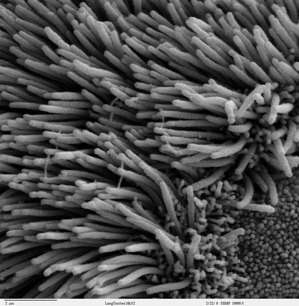
Dust Mop
This photo (Figure 7.3.1) looks like a close-up of an old fashioned dust mop, and the object it shows has a somewhat similar function. The object, however, is greatly enlarged in the photo. Can you guess what it is? The answer may surprise you.
It is a scanning-electron micrograph of human epithelial cells that line the bronchial passages. The floppy, dust-mop-like extensions are actually microscopic structures called cilia projecting from the outer surface of the epithelial cells. The function of the cilia is to trap dust, pathogens, and other particles in the air before it enters the lungs. The cilia also sway back and forth to sweep the trapped particles upward toward the throat, from which they can be expelled from the body.
Human Cells
Like the ciliated bronchial cells in the micrograph above, many other cells in the human body are very distinctive and well-suited for special functions. To perform their special functions, cells may vary in a number of ways.
Variation in Human Cells
Some cells act as individual cells and are not attached to one another. Red blood cells are a good example. Their main function is to transport oxygen to other cells throughout the body, so they must be able to move freely through the circulatory system. Many other cells, in contrast, act together with other similar cells as part of the same tissue, so they are attached to one another and cannot move freely. For example, epithelial cells lining the respiratory tract are attached to each other to form a continuous surface that protects the respiratory system from particles and other hazards in the air.
Many cells can divide readily and form new cells. Skin cells are constantly dying and being shed from the body and replaced by new skin cells, and bone cells can divide to form new bone for growth or repair. On the other hand, some other cells — like certain nerve cells — can divide and form new cells only under exceptional circumstances. Nervous system injuries (such as a severed spinal cord) generally cannot heal by the production of new cells, which results in a permanent loss of function.
Many human cells have the primary job of producing and secreting a particular substance, such as a hormone or an enzyme. For example, special cells in the pancreas produce and secrete the hormone insulin, which regulates the level of glucose in the blood. Some of the epithelial cells that line the bronchial passages produce mucus, a sticky substance that helps trap particles in the air before it passes into the lungs.
Different, but Identical
All the different cell types within an individual human organism are genetically identical, so no matter how different the cells are, they all have the same genes. How can such different types of cells arise? The answer is differential regulation of genes. Cells with the same genes can be very different because different genes are expressed depending on the cell type.
Examples of Human Cell Types
Many common types of human cells — such as bone cells and white blood cells — actually consist of several subtypes of cells. Each subtype, in turn, has a special structure and function. A closer look at these cell types will give you a better appreciation for the diversity of structures and functions of human cells.
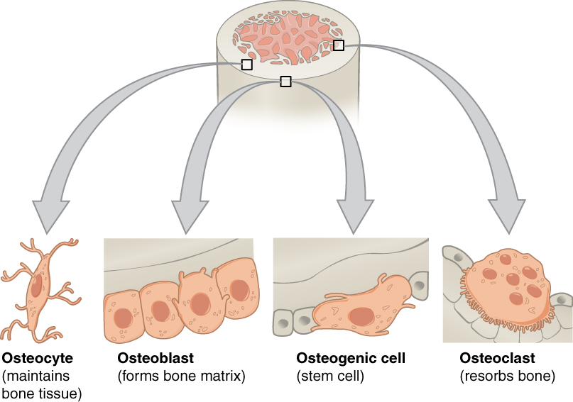
Bone Cells
There are four main subtypes of bone cells, as shown in Figure 7.3.2. Each type has a different form and function:
- Osteogenic cells are undifferentiated stem cells that differentiate to form osteoblasts in the tissue that covers the outside of bone.
- Osteoblasts are immature bone cells that are involved in synthesizing new bone. They develop into osteocytes, or mature bone cells.
- Osteocytes are star-shaped bone cells that make up the majority of bone tissue. They are the most common cells in mature bone.
- Osteoclasts are very large, multinucleated cells responsible for the breakdown of bones through resorption. The breakdown of bone is very important in bone health, because it allows for bone remodeling.
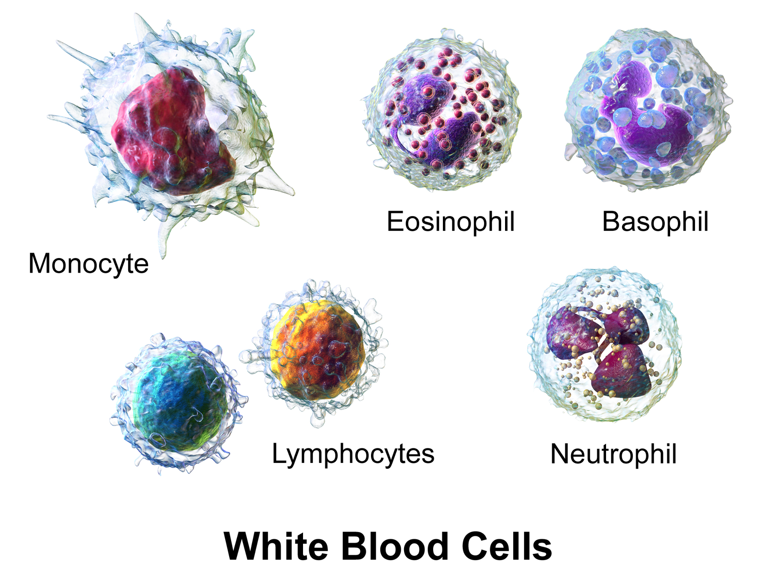
White Blood Cells
White blood cells (also called leukocytes) are even more variable than bone cells. Five subtypes of white blood cells are shown in Figure 7.3.3. All of them are immune system cells involved in defending the body, but each subtype has a different function. They also differ in the normal proportion of all leukocytes they make up.
- Monocytes make up about five per cent of leukocytes. They engulf and destroy (phagocytize) pathogens in tissues.
- Eosinophils compose about two per cent of leukocytes. They attack larger parasites and set off allergic responses.
- Basophils make up less than one per cent of leukocytes. They release proteins called histamines that are involved in inflammation.
- Lymphocytes make up about 30 per cent of leukocytes. They include B cells and T cells. B cells produce antibodies against nonself antigens, and T cells destroy virus-infected cells and cancer cells.
- Neutrophils are the most numerous white blood cells, making up about 62 per cent of leukocytes. They phagocytize single-celled bacteria and fungi in the blood.
Tissues
Groups of connected cells form tissues. The cells in a tissue may all be the same type, or they may be of multiple types. In either case, the cells in the tissue work together to carry out a specific function. There are four main types of human tissues: connective, epithelial, muscle, and nervous tissues. We will be learning about each of the four tissue types in more detail in the next few sections, but here is a summary:
Epithelial Tissue
Epithelial tissue is made up of cells that line inner and outer body surfaces, such as the skin and the inner surface of the digestive tract. Epithelial tissue that lines inner body surfaces and body openings is called mucous membrane. This type of epithelial tissue produces mucus, a slimy substance that coats mucous membranes and traps pathogens, particles, and debris. Epithelial tissue protects the body and its internal organs, secretes substances (such as hormones) in addition to mucus, and absorbs substances (such as nutrients).
Epithelial tissue is identified and named by shape and layering. Epithelial cells exist in three main shapes: squamous, cuboidal, and columnar. These specifically-shaped cells can, depending on function, be layered several different ways: simple, stratified, pseudostratified, and transitional.
Connective Tissue
Bone and blood are examples of connective tissue. Connective tissue is very diverse. In general, it forms a framework and support structure for body tissues and organs. It’s made up of living cells separated by non-living material, called an extracellular matrix, which can be solid or liquid. The extracellular matrix of bone, for example, is a rigid mineral framework. The extracellular matrix of blood is liquid plasma. Connective tissues such as bone and cartilage generally form the body’s structure.
There are three main categories of connective tissue, based on the nature of the matrix. They look very different from one another, which is a reflection of their different functions:
- Fibrous connective tissue: is characterized by a matrix which is flexible and is made of protein fibres including collagen, elastin and possibly reticular fibres. These tissues are found making up tendons, ligaments, and body membranes.
- Supportive connective tissue: is characterized by a solid matrix and is what is used to make bone and cartilage. These tissues are used for support and protection.
- Fluid connective tissue: is characterized by a fluid matrix and includes both blood and lymph.
Muscular Tissue

Muscular tissue is made up of cells that have the unique ability to contract. There are three major types of muscle tissue, as pictured in Figure 7.3.4: skeletal, smooth, and cardiac muscle tissues.
- Skeletal muscles are striated (or striped) in appearance, because of their internal structure. Skeletal muscles are attached to bones by tendons, a type of connective tissue. When they pull on the bones, they enable the body to move. Skeletal muscles are under voluntary control.
- Smooth muscles are nonstriated muscles. They are found in the walls of blood vessels and in the reproductive, gastrointestinal, and respiratory tracts. Smooth muscles are not under voluntary control.
- Cardiac muscles are striated and found only in the heart. Their contractions cause the heart to pump blood. Cardiac muscles are not under voluntary control.
Nervous Tissue
Nervous tissue is made up of neurons and other types of cells, generally called glial cells. Neurons transmit messages — usually through an electrochemical process — with support from the other cells. Nervous tissue makes up the central nervous system (mainly the brain and spinal cord) and peripheral nervous system (the network of nerves that runs throughout the rest of the body).
Feature: My Human Body
If you are a blood donor, then you have donated tissue. Blood is a tissue that you can donate when you are alive. You may have indicated on your driver’s license application that you wish to be a tissue donor in the event of your death. Deceased people can donate many different tissues, including skin, bone, heart valves, and the corneas of the eyes. If you are not already registered as a tissue donor, the information from the BC Transplant link below may help you decide if you would like to register.
As of April 2020, there were over 1.5 million donors registered in the BC Organ Donor Registry. In 2019, 480 lives were saved in BC as a result of organ donation, both through deceased donors and living donors. Over the years, organ transplants have saved the lives of over 5,000 British Columbians. In 2019, 331 kidney transplants were conducted, the most common transplant needed, and of these, 120 kidneys were from living donors — people who donated their kidney and are still walking around to tell the tale! Despite these encouraging statistics, there are still over 640 British Columbians on the waiting list for a kidney transplant.
Unlike organs — which generally must be transplanted immediately after the donor dies — donated tissues can be processed and stored for a long time for later use. Donated tissues can be used to replace burned skin and damaged bone, and to repair ligaments. Corneal tissues can be used for corneal transplants that restore sight in blind people. Unfortunately, there are not enough tissues to go around, and the need for donated tissues keeps rising.
For more information on organ and tissue transplants, you can visit the BC Transplant website.
7.3 Summary
- Cells of the human body show a lot of variation. Some cells are unattached to other cells and can move freely, while others are attached to each other and cannot move freely. Some cells can divide readily and form new cells, and others can divide only under exceptional circumstances. Many cells are specialized to produce and secrete particular substances.
- All the different cell types within an individual have the same genes. Cells can vary because different genes are expressed depending on the cell type.
- Many common types of human cells actually consist of several subtypes of cells, each of which has a special structure and function. Subtypes of bone cells, for example, include osteocytes, osteoblasts, osteogenic cells, and osteoclasts.
- There are four major types of human tissues: connective, epithelial, muscle, and nervous tissues.
- Connective tissues, such as bone and blood, are made up of living cells that are separated by non-living material, called extracellular matrix.
- Epithelial tissues, such as skin and mucous membranes, protect the body and its internal organs. They also secrete or absorb substances.
- Muscle tissues are made up of cells that have the unique ability to contract. They include skeletal, smooth, and cardiac muscle tissues.
- Nervous tissues are made up of neurons — which transmit messages — and glial cells of various types, which play supporting roles.
7.3 Review Questions
- Give an example of cells that function individually and move freely. Additionally, give an example of cells that act together and are attached to other cells of the same type.
- What is an example of cells that can readily divide? What is an example of cells that can divide only under rare circumstances?
- Identify a type of cell that secretes an important substance. Name the substance it secretes.
- Explain how different cell types come about when all the cells in an individual human being are genetically identical.
- Compare and contrast four subtypes of human bone cells.
- Identify three types of human white blood cells. State their functions.
- Why are bone and blood both classified as connective tissues?
- Name another type of connective tissue. Describe its role in the human body.
- Based on the information above about types of epithelial tissues, list four general ways this type of tissue functions in the human body.
- Compare and contrast the three types of muscle tissues.
- Identify the two main types of cells that make up nervous tissue. Compare their general functions.
- Of the main types of human tissue, name two that can secrete hormones.
- Cells in a particular tissue…
- Are all of the same type
- Have different genes from cells in other tissues
- Work together to carry out a function
- Are always connected physically to each other
- Why are mucus membranes often located in regions that interface between the body and the outside world?
- Skin is a type of _____________ tissue.
- Body fat is a type of ____________ tissue.
7.3 Explore More
Could tissue engineering mean personalized medicine? – Nina Tandon, TED-Ed, 2013.
Printing a human kidney – Anthony Atala, TED-Ed, 2013.
Attributions
Figure 7.3.1
Bronchiolar_epithelium_4_-_SEM by Louisa Howard on Wikimedia Commons is released into the public domain (https://en.wikipedia.org/wiki/Public_domain).
Figure 7.3.2
Bone_cells by OpenStax College on Wikimedia Commons is used under a CC BY 3.0 (https://creativecommons.org/licenses/by/3.0/deed.en) license.
Figure 7.3.3
- Blausen_0909_WhiteBloodCells by BruceBlaus on Wikimedia Commons is used under a CC BY 3.0 (https://creativecommons.org/licenses/by/3.0) license.
- 414 Skeletal Smooth Cardiac by OpenStax College on Wikimedia Commons is used under a CC BY 3.0 (https://creativecommons.org/licenses/by/3.0/deed.en) license.
References
Betts, J. G., Young, K.A., Wise, J.A., Johnson, E., Poe, B., Kruse, D.H., Korol, O., Johnson, J.E., Womble, M., DeSaix, P. (2013, April 25). Figure 4.18 Muscle tissue [digital image]. In Anatomy and Physiology (Section 4.4). OpenStax. https://openstax.org/books/anatomy-and-physiology/pages/4-4-muscle-tissue-and-motion
Betts, J. G., Young, K.A., Wise, J.A., Johnson, E., Poe, B., Kruse, D.H., Korol, O., Johnson, J.E., Womble, M., DeSaix, P. (2013, April 25). Figure 6.11 Bone cells [digital image]. In Anatomy and Physiology (Section 6.3). OpenStax. https://openstax.org/books/anatomy-and-physiology/pages/6-3-bone-structure
Blausen.com Staff. (2014). Medical gallery of Blausen Medical 2014. WikiJournal of Medicine 1 (2). DOI:10.15347/wjm/2014.010. ISSN 2002-4436.
Brainard, J/ CK-12 Foundation. (2016). Figure 3 Five subtypes of human white blood cells in the human immune system [digital image]. In CK-12 College Human Biology (Section 9.3) [online Flexbook]. CK12.org. https://www.ck12.org/book/ck-12-college-human-biology/section/9.3/
TED-Ed. (2013, May 17). Could tissue engineering mean personalized medicine? – Nina Tandon. YouTube. https://www.youtube.com/watch?v=_7TKiFRkKGY&feature=youtu.be
TED-Ed. (2013, March 15). Printing a human kidney – Anthony Atala. YouTube. https://www.youtube.com/watch?v=bX3C201O4MA&feature=youtu.be
Tissue which lines the outer surfaces of organs and blood vessels throughout the body, as well as the inner surfaces of cavities in many internal organs. An example is the epidermis, the outermost layer of the skin. There are three principal shapes of epithelial cell: squamous, columnar, and cuboidal.
One of the four basic types of tissue, connective tissue is found in between other tissues everywhere in the body, including the nervous system and generally forms a framework and support structure for body tissues and organs.
A three-dimensional network of extracellular macromolecules, such as collagen, enzymes, and glycoproteins, that provide structural and biochemical support to surrounding cells.
A soft tissue that composes muscles in animal bodies, and gives rise to muscles' ability to contract. This is opposed to other components or tissues in muscle such as tendons or perimysium.
A specialized tissue found in the central nervous system and the peripheral nervous system. It consists of neurons and supporting cells called neuroglia. The nervous system is responsible for the control of the body and the communication among its parts.
A person who donates one kidney or a portion of their liver or a part of their lung to someone who needs those organs to survive. Transplants from living donors have been extremely successful and most donors recover with very few complications.
