16.6 Disorders of the Urinary System
Created by CK-12 Foundation/Adapted by Christine Miller

Awareness Ribbon
Awareness ribbons are symbols meant to show support or to raise consciousness for a cause. Different colours are associated with different issues, often relating to health problems. The first ribbon to gain familiarity for a health issue was the red ribbon for HIV/AIDS, created in 1991. The pink ribbon for breast cancer awareness is probably the best known today. Do you know what a green ribbon like the one pictured in Figure 16.6.1 represents? Among several other health problems, a green ribbon is meant to show support or raise awareness for kidney disorders.
Disorders of the Kidneys
The kidneys play such vital roles in eliminating wastes and toxins — and in maintaining body-wide homeostasis — that disorders of the kidneys may be life threatening. Gradual loss of normal kidney function commonly occurs with a number of disorders, including diabetes mellitus and high blood pressure. Other disorders of the kidneys are caused by faulty inherited genes. Loss of kidney function may eventually progress to kidney failure.
Diabetic Nephropathy
Diabetic nephropathy is a progressive kidney disease caused by damage to the capillaries in the glomeruli of the kidneys, due to long-standing diabetes mellitus (see Figure 16.6.2). It is not fully understood how diabetes leads to damage of glomerular capillaries, but it is thought that high levels of glucose in the blood are involved. In people with diabetes, diabetic nephropathy is more likely if blood glucose is poorly controlled. Having high blood pressure, a history of cigarette smoking, and a family history of kidney problems are additional risk factors. Diabetic nephropathy often has no symptoms at first. In fact, it may take up to a decade after kidney damage begins for symptoms to appear. When they do appear, they typically include severe tiredness, headaches, nausea, frequent urination, and itchy skin.
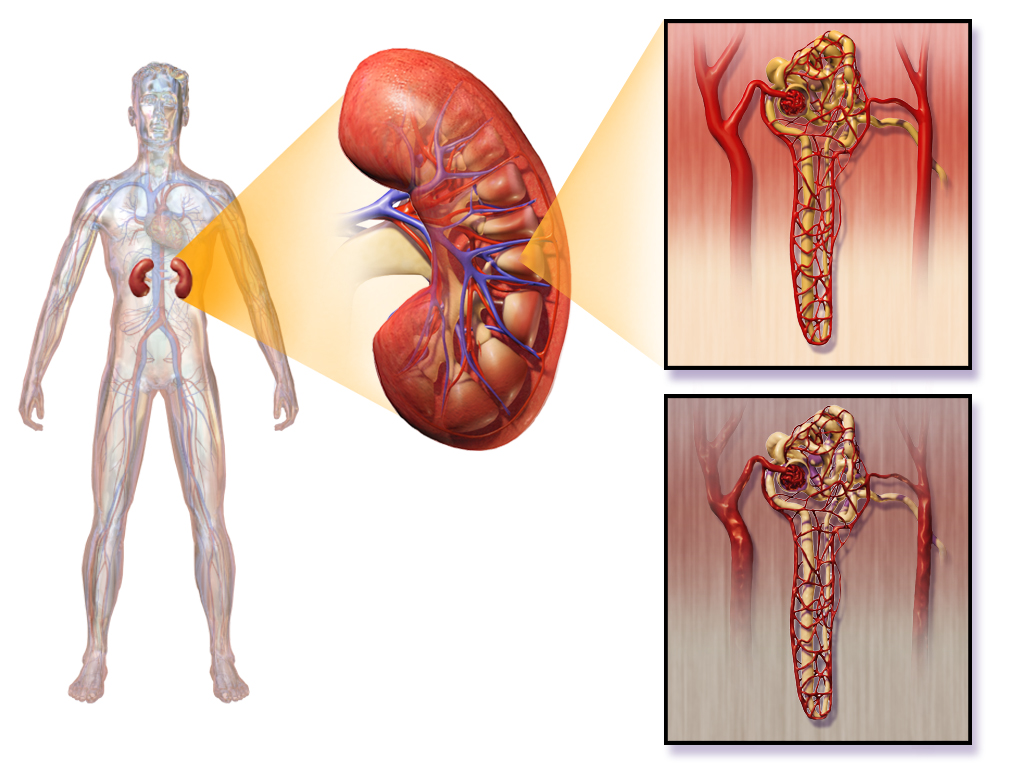
Proteins are large molecules that are usually not filtered out of blood in the glomeruli. When the glomerular capillaries are damaged, it allows proteins (such as albumin) to leak into the filtrate from the blood. As a result, albumin ends up being excreted in the urine. Finding a high level of albumin in the urine is one indicator of diabetic nephropathy and helps to diagnose the disorder. Drugs may be prescribed to reduce protein levels in the urine. Controlling high blood sugar levels and hypertension (high blood pressure) is also important to help slow kidney damage, as is a reduction of sodium intake.
Polycystic Kidney Disease
Polycystic kidney disease (PKD) is a genetic disorder in which multiple abnormal cysts develop and grow in the kidneys. Figure 16.6.3 shows a pair of kidneys that are riddled with cysts from PKD. In people who inherit PKD, the cysts may start to form at any point in life, from infancy through adulthood. Typically, both kidneys are affected. Symptoms of the disorder may include high blood pressure, headaches, abdominal pain, blood in the urine, and excessive urination.
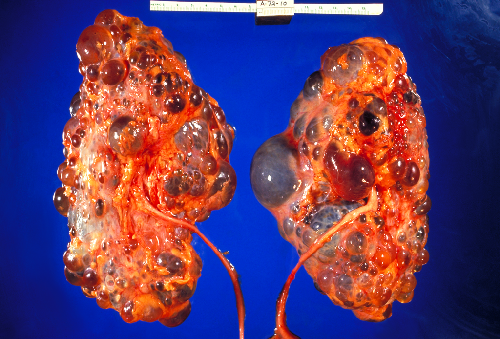
There are two types of PKD. The more common type is caused by an autosomal dominant allele, and the less common type is caused by an autosomal recessive allele. Both types together make PKD one of the most common hereditary diseases in Canada, affecting one in every 500 people. There is little or no difference in the rate of occurrence of PKD between genders or ethnic groups. Other than a kidney transplant, there is no known cure for this disease.
Kidney Failure
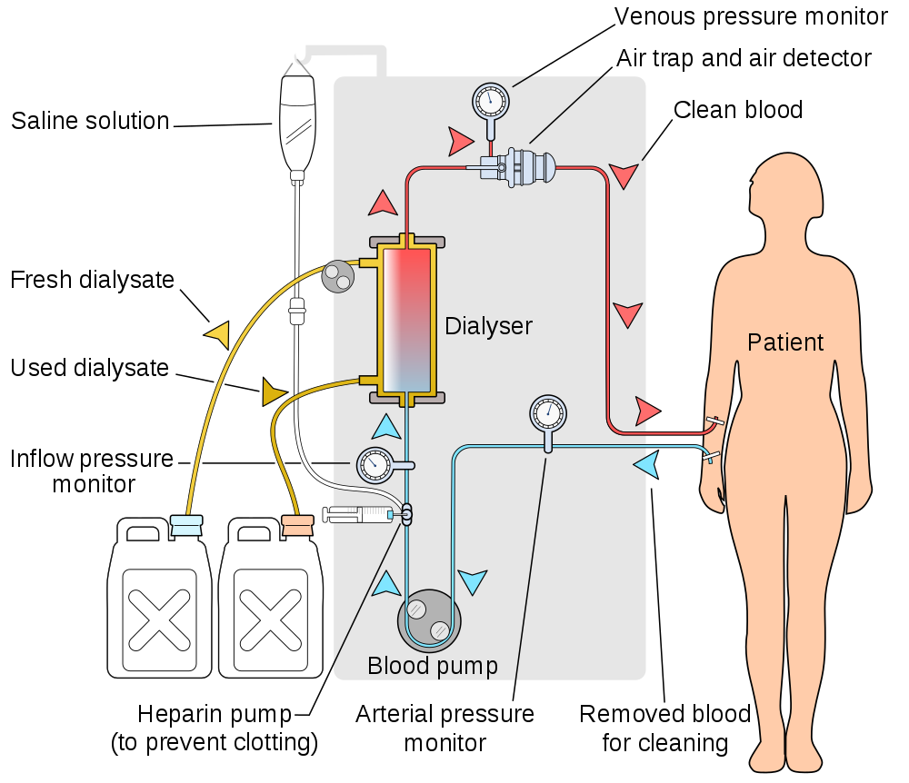
Both diabetic nephropathy and PKD may lead to kidney (or renal) failure(classified as end-stage kidney disease), in which the kidneys are no longer able to adequately filter metabolic wastes from the blood. Long-term, uncontrolled high blood pressure is another common cause of kidney failure. Symptoms of kidney failure may include nausea, more or less frequent urination, blood in the urine, muscle cramps, anemia, swelling of the extremities, and shortness of breath due to the accumulation of fluid in the lungs. If kidney function drops below the level needed to sustain life, then the only treatment option is kidney transplantation or some means of artificial filtration of the blood, such as by hemodialysis.
Hemodialysis is a medical procedure in which blood is filtered externally through a machine. You can see how it works in Figure 16.6.4. During dialysis, waste products (such as urea) are removed — along with excess water — from the patient’s blood before the blood is returned to the patient. Hemodialysis is typically done on an outpatient basis in a hospital or special dialysis clinic. Less frequently, it is done in the patient’s home. Depending on the patient’s size, among other factors, the blood is filtered for three to four hours roughly three times a week.
Kidney Stones
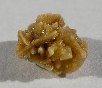
A kidney stone, (pictured in Figure 16.6.5) also known as a renal calculus, is a solid crystal that forms in a kidney from minerals in urine (see Figure 16.6.6). The majority of kidney stones consist of crystals of calcium salts. Kidney stones typically leave the body in the urine stream. A small stone may go undetected, because it can pass through the ureters and other urinary tract organs without causing symptoms. A larger stone may cause pain when it passes through the urinary tract. If a kidney stone grows large enough, it may block the ureter. Blockage of a ureter may cause a decrease in kidney function and damage to the kidney.
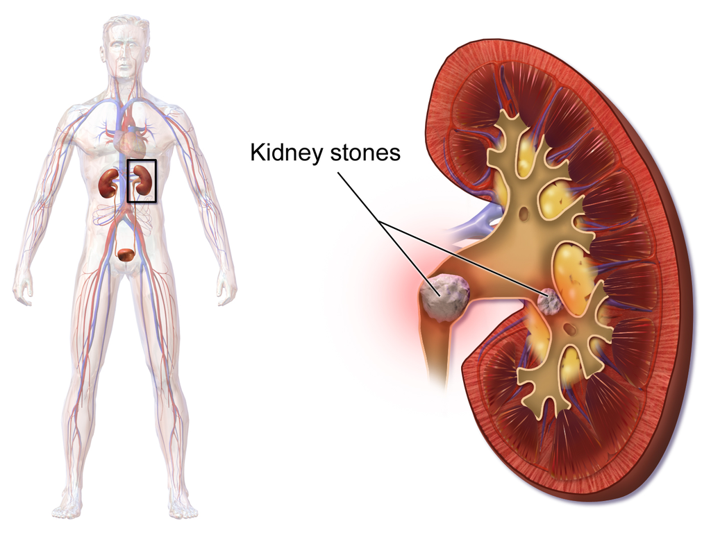
A kidney stone that causes pain is generally treated with pain medication, such as opiates, until it passes through the urinary tract. A stone that causes a blockage may be treated with lithotripsy. This is a medical procedure in which high-intensity ultrasound pulses are applied externally to cause fragmentation of the stone into pieces small enough to pass easily through the urinary tract. Although lithotripsy is noninvasive, it can cause damage to the kidneys. An alternative treatment for a stone that blocks urine flow is to insert a stent into the ureter to expand it and allow both urine and the stone to pass. In some cases, surgery may be required to physically remove a large stone from the ureter. In minor cases, sometimes drinking apple cider vinegar or lemon juice can break down small kidney stones because of the citric acid these foods contain.
A combination of lifestyle and genetic factors seem to predispose certain people to develop kidneys stones. Risk factors include high consumption of cola soft drinks, eating a diet high in animal protein, being overweight, and not drinking enough fluids. Preventive measures are obvious. They include limiting cola consumption, eating less animal protein, losing weight, and increasing fluid intake.
Other Urinary System Disorders
Although disorders of the kidneys are generally the most serious urinary system disorders, problems that affect other organs of the urinary tract are generally more common. They include bladder infections and urinary incontinence.
Bladder Infection
A bladder infection, also called cystitis, is a very common type of urinary tract infection in which the urinary bladder becomes infected by bacteria (typically E. coli), and rarely by fungi. Symptoms of bladder infections may include pain with urination, frequent urination, and feeling the need to urinate despite having an empty bladder. In some cases, there may be blood in the urine. A much less common type of urinary tract infection is pyelonephritis, in which the kidney becomes infected. If a kidney infection occurs, it is generally because of an untreated bladder infection. Bladder infections are treated mainly with antibiotics.
Risk factors for urinary bladder infections include sexual intercourse, improper toileting technique, diabetes, obesity, and — most notably — female sex. Bladder infections are four times more common in women than in men. For women, they are the most common type of bacterial infections, and as many as one in ten women have a bladder infection in any given year. Female anatomy explains the sex difference in the incidence of bladder infections. The urethra is much shorter and closer to the anus in females than in males, so contamination of the urethra and then the bladder with GI tract bacteria is more likely in females than in males. Once the bacteria reach the bladder, they can attach to the bladder wall and form a biofilm that resists the body’s immune response.
Urinary Incontinence
Urinary incontinence is a chronic problem of uncontrolled leakage of urine. It is very common, especially at older ages, and especially in women. Sometimes, urinary incontinence is a sign of another health problem, such as diabetes or obesity. Regardless of the underlying cause, the symptoms of urinary incontinence alone may have a large impact on quality of life, frequently causing inconvenience, embarrassment, and distress.
In men, urinary incontinence is most commonly caused by an enlarged prostate gland or treatment for prostate cancer. In women, there are two common types of urinary incontinence with different causes: stress incontinence and urge incontinence.
- Stress urinary incontinence is caused by loss of support of the urethra, usually due to stretching of pelvic floor muscles during childbirth. It is characterized by leakage of small amounts of urine with activities that increase abdominal pressure, such as coughing, sneezing, or lifting. Treatment of stress urinary incontinence may include Kegel exercises to strengthen the pelvic muscles. More serious cases may call for surgery to improve support for the bladder.
- Urge urinary incontinence (commonly called “overactive bladder”) is caused by uncontrolled contractions of the detrusor muscle in the wall of the bladder. This causes the bladder to empty unexpectedly. Urge incontinence is characterized by leakage of large amounts of urine in association with insufficient warning to get to the bathroom in time. Treatment of urge incontinence may include taking a medication to relax the detrusor muscle.
Feature: My Human Body
You probably have had to “donate” a urine specimen for analysis in conjunction with a medical visit. A thorough medical exam often includes clinical tests for urine. Understanding what your urine can reveal about your health may help you appreciate the need for such tests.
The most common urine test is called urinalysis. In a routine urinalysis, a urine sample may be analyzed by sight and smell, and with simple urine test strips. If a particular disorder is suspected, urinalysis may be more extensive. The urine may be analyzed with specific tests or viewed under a microscope to identify abnormal substances in the urine. If a bacterial infection is suspected, a sample of urine may be cultured in the lab to see if it grows bacteria, and which type of bacteria grow. Knowing the type of bacteria is important for deciding which class of antibiotics is likely to be most effective in treating the infection.
The colour and clarity of urine may be obvious first indicators of disorders or other abnormalities. Normal urine is yellow to amber in colour, and looks clear. If urine is nearly colourless, it could be a sign of excessive fluid intake, or it might be a sign of diabetes. Very dark urine may indicate dehydration, but it could also be caused by taking certain medications or ingesting some other substances. If urine has a reddish tinge, it is often a sign of blood in the urine, which could be due to a urinary tract infection, kidney stone, or even cancer. If urine appears cloudy instead of clear, it could be due to white blood cells in the urine, which may be another sign of a urinary tract infection.
If it is very diluted, normal urine may have virtually no odor. It will have a stronger odor if it is concentrated. Brief changes in the normal odor of urine often occur due to the ingestion of certain foods or medications. After eating asparagus, for example, urine may have a peculiar and distinctive odor for several hours. More significant is urine that has a sweet smell, because this may indicate sugar in the urine, which is a sign of diabetes.
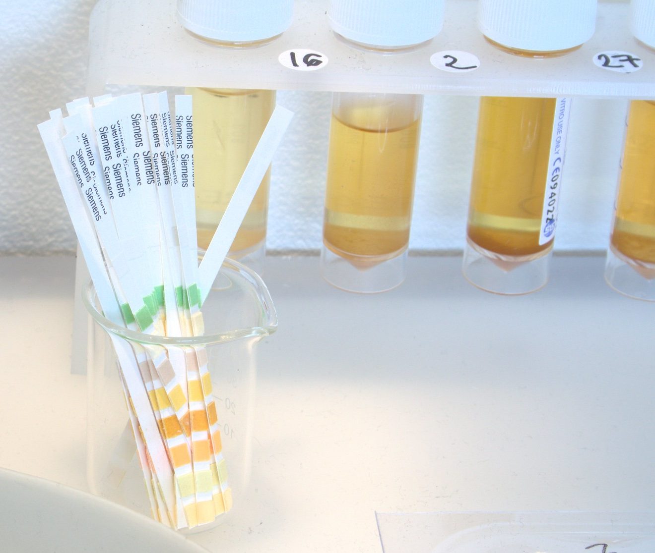
Urine test strips (shown in Figure 16.6.7), much like the familiar litmus test strips used to detect acids and bases in chemistry lab, are used to identify abnormal levels of certain components in the urine. For example, urine test strips can detect and quantify the presence of nitrites in urine, which is usually a sign of infection with certain types of bacteria. Urine test strips can also be used to identify proteins such as albumin in urine, which may be a sign of a kidney infection or of kidney failure. Levels of sodium in urine can also be measured with test strips, and higher-than-normal levels may be another indication of kidney failure. In addition, test strips can identify and quantify the presence of white blood cells and blood in a urine specimen, both of which are likely to be a sign of a urinary tract infection or some other urinary system disorder.
Besides the use of urine test strips, other simple urine tests that are often performed include Benedict’s test, which is a test for the presence and quantity of glucose in urine. If the level is high, it likely indicates diabetes. The test is so simple that it may even be done by the patient at home to monitor how well sugar levels are being controlled. Testing for some other substances in urine requires the patient to collect urine over a 24-hour period. This is the case when testing for the adrenal hormone cortisol. When urine cortisol levels are higher than normal, it may indicate Cushing’s syndrome. When the levels are lower than normal, it may indicate Addison’s disease.
16.6 Summary
- Diabetic nephropathy is a progressive kidney disease caused by damage to the capillaries in the glomeruli of the kidneys due to long-standing diabetes mellitus. Years of capillary damage may occur before symptoms first appear.
- Polycystic kidney disease (PKD) is a genetic disorder (autosomal dominant or recessive) in which multiple abnormal cysts grow in the kidneys.
- Diabetic nephropathy, PKD, or chronic hypertension may lead to kidney failure, in which the kidneys are no longer able to adequately filter metabolic wastes from the blood. Kidneys may fail to such a degree that kidney transplantation or repeated, frequent hemodialysis is needed to support life. In hemodialysis, the patient’s blood is filtered artificially through a machine, and then returned to the patient’s circulation.
- A kidney stone is a solid crystal that forms in a kidney from minerals in urine. A small stone may pass undetected through the ureters and the rest of the urinary tract. A larger stone may cause pain when it passes, or be too large to pass, causing blockage of a ureter. Large kidney stones may be shattered with high-intensity ultrasound into pieces small enough to pass through the urinary tract, or they may be removed surgically.
- A bladder infection is generally caused by bacteria that reach the bladder from the GI tract and multiply. Bladder infections are much more common in females than males, because the female urethra is much shorter and closer to the anus. Treatment generally includes antibiotic drugs.
- Urinary incontinence is a chronic problem of uncontrolled leakage of urine. It is very common, especially at older ages and in women. In men, urinary incontinence is usually caused by an enlarged prostate gland. In women, it is usually caused by stretching of pelvic floor muscles during childbirth (stress incontinence) or by an “overactive bladder” that empties without warning (urge incontinence).
16.6 Review Questions
-
- Define kidney failure.
- When kidney function drops below the level needed to sustain life, what are potential treatments for kidney failure?
- Describe hemodialysis.
- How may a large kidney stone be removed from the body?
- How are bladder infections usually treated?
- Why are bladder infections much more common in females than in males?
- Compare and contrast stress incontinence and urge incontinence.
- Why is the presence of a protein(such as albumin) in the urine a cause for concern?
- Patients undergoing hemodialysis usually have to do this procedure a few times a week. Why does it need to be done so frequently?
16.6 Explore More
Urinary Tract Infections, Animation, Alila Medical Media, 2016.
What causes kidney stones? – Arash Shadman, TED-Ed, 2017.
Kegel Exercises Beginners Workout For Women, Michelle Kenway, 2013.
Attributions
Figure 16.6.1
512px-Green_ribbon.svg by MesserWoland on Wikimedia Commons is used under a CC BY-SA 3.0 (http://creativecommons.org/licenses/by-sa/3.0/) license.
Figure 16.6.2
1024px-Blausen_0310_DiabeticNephropathy by BruceBlaus on Wikimedia Commons is used under a CC BY 3.0 (https://creativecommons.org/licenses/by/3.0) license.
Figure 16.6.3
1024px-Polycystic_kidneys,_gross_pathology_CDC_PHIL by Dr. Edwin P. Ewing, Jr. / CDC‘s Public Health Image Library (PHIL) #861. on Wikimedia Commons is in the public domain (https://en.wikipedia.org/wiki/Public_domain).
Figure 16.6.4
1000px-Hemodialysis-en.svg by User:YassineMrabet on Wikimedia Commons is used under a CC BY 3.0 (https://creativecommons.org/licenses/by/3.0) license.
Figure 16.6.5
512px-Kidney_stone_1 by User:Михајло Анђелковић on Wikimedia Commons is used under a CC BY-SA 3.0 (https://creativecommons.org/licenses/by-sa/3.0) license.
Figure 16.6.6
Blausen_0595_KidneyStones by BruceBlaus on Wikimedia Commons is used under a CC BY 3.0 (https://creativecommons.org/licenses/by/3.0) license.
Figure 16.6.7
Amanda Cotton – Urinalysis Test Strips by Dominic Alves on Flickr is used under a CC BY 2,0 (https://creativecommons.org/licenses/by/2.0/) license.
References
Alila Medical Media. (2016, September 8). Urinary tract infections, animation. YouTube. https://www.youtube.com/watch?v=lY2bZjggc08&feature=youtu.be
Blausen.com Staff. (2014). Medical gallery of Blausen Medical 2014. WikiJournal of Medicine 1 (2). DOI:10.15347/wjm/2014.010. ISSN 2002-4436.
Michelle Kenway. (2013, February 1). Kegel exercises beginners workout for women. YouTube. https://www.youtube.com/watch?v=wRKhtfbJHdo&feature=youtu.be
TED-Ed. (2017, July 3). What causes kidney stones? – Arash Shadman. YouTube. https://www.youtube.com/watch?v=W0GpIMNTPYg&feature=youtu.be
Updated Canadian expert consensus published to guide optimal management of ADPKD. (2018, December 18). PDK Foundation of Canada. https://www.endpkd.ca/canadian_expert_consensus_2018
One of a pair of organs of the excretory and urinary systems that filters wastes and excess water out of blood and forms urine.
The ability of an organism to maintain constant internal conditions despite external changes.
A disorder in which blood sugar (glucose) levels are abnormally high because the body does not produce enough insulin to meet its needs.
The measure of the force exerted by circulating blood on the walls of arteries.
A progressive kidney disease caused by damage to capillaries in the glomeruli of the kidneys due to poor blood sugar control in people with diabetes.
Glucose (also called dextrose) is a simple sugar with the molecular formula C6H12O6. Glucose is the most abundant monosaccharide, a subcategory of carbohydrates. Glucose is mainly made by plants and most algae during photosynthesis from water and carbon dioxide, using energy from sunlight.
A genetic disorder in which multiple abnormal cysts develop and grow in the kidneys.
Any chromosome that is not a sex chromosome.
Refers to the relationship between two versions of a gene. Individuals receive two versions of each gene, known as alleles, from each parent. If the alleles of a gene are different, one allele will be expressed; it is the dominant gene. The effect of the other allele, called recessive, is masked.
A variant form of a given gene, meaning it is one of two or more versions of a known mutation at the same place on a chromosome. It can also refer to different sequence variations for a several-hundred base-pair or more region of the genome that codes for a protein.
A gene that can be masked by a dominant gene. In order to have a trait that is expressed by a recessive gene, such as blue eyes, you must get the gene for blue eyes from both of your parents.
The loss of the ability of nephrons in the kidney to function fully due to a progressive kidney disease such as diabetic nephropathy or polycystic kidney disease.
A medical procedure for patients with kidney failure in which wastes and excess water are artificially filtered out of blood by passing it through a machine.
A solid crystal that forms in a kidney from minerals such as calcium in urine.
A common type of urinary tract infection in which the bladder becomes infected, usually by bacteria but occasionally by fungi.
An inflammation of the substance of the kidney as a result of bacterial infection.
A common chronic problem of uncontrolled leakage of urine.
The unintentional loss of urine. Stress incontinence happens when physical movement or activity — such as coughing, laughing, sneezing, running or heavy lifting — puts pressure (stress) on your bladder, causing you to leak urine.
When you have a strong, sudden need to urinate that is difficult to delay. The bladder then squeezes, or spasms, and you lose urine.
An analysis of urine by physical, chemical, and microscopical means to test for the presence of disease, drugs, etc.
A disease caused by problems with the pancreatic hormone insulin, which leads to high blood glucose levels and symptoms such as excessive thirst and urination; includes type 1 and type 2 diabetes.
A disorder in which there is hypersecretion of the adrenal cortex hormone cortisol, most commonly due to a tumor of the pituitary gland.
A disorder characterized by hyposecretion of the adrenal cortex hormone cortisol, generally because the immune system attacks and destroys the adrenal gland.
A network of capillaries in the nephron of a kidney where substances are filtered out of the blood.
