15.4 Upper Gastrointestinal Tract
Created by CK-12 Foundation/Adapted by Christine Miller

Head Stand
Did you ever wonder what would happen if you tried to swallow food while standing on your head like this person in Figure 15.4.1? Many people think that food travels down the gullet from the mouth by the force of gravity. If that were the case, then food you swallowed would stay in your throat while you were standing on your head. In reality, your position doesn’t have much to do with your ability to swallow. Food will travel from your mouth to your stomach whether you are standing upright or upside down. That’s because the tube the food travels through — the esophagus — moves the food along via muscular contractions known as peristalsis. The esophagus is one of several organs that make up the upper gastrointestinal tract.
Organs of the Upper Gastrointestinal Tract
Besides the esophagus, organs of the upper gastrointestinal (GI) tract include the mouth, pharynx, and stomach. These hollow organs are all connected to form a tube through which food passes during digestion. The only role in digestion played by the pharynx and esophagus is to move food through the GI tract. The mouth and stomach, in contrast, are organs where digestion — or the breakdown of food — also occurs. In both of these organs, food is broken into smaller pieces (mechanical digestion), as well as broken down chemically (chemical digestion). It should be noted that the first part of the small intestine (duodenum) is considered in some contexts to be part of the upper GI tract, but that practice is not followed here.
Mouth
The mouth is the first organ of the GI tract. Most of the oral cavity is lined with mucous membrane. This tissue produces mucus, which helps moisten, soften, and lubricate food. Underlying the mucous membrane is a thin layer of smooth muscle to which the mucous membrane is only loosely connected. This gives the mucous membrane considerable ability to stretch as you eat food. The roof of the mouth, called the palate, separates the oral cavity from the nasal cavity. The front part is hard, consisting of mucous membrane covering a plate of bone. The back part of the palate is softer and more pliable, consisting of mucous membrane over muscle and connective tissue. The hard surface of the front of the palate allows for pressure needed in chewing and mixing food. The soft, pliable surface of the back of the palate can move to accommodate the passage of food while swallowing. Muscles at either side of the soft palate contract to create the swallowing action.
Several specific structures in the mouth are specialized for digestion. These include salivary glands, tongue, and teeth.
Salivary Glands
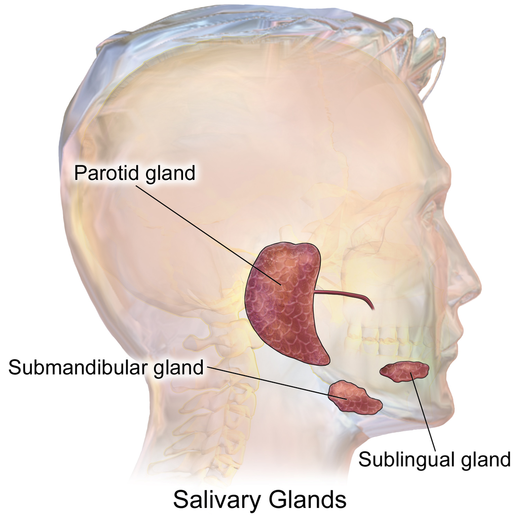
The mouth contains three pairs of major salivary glands, shown in Figure 15.4.2. These three pairs are all exocrine glands that secrete saliva into the mouth through ducts.
- The largest of the three major pairs of salivary glands are the parotid glands, which are located on either side of the mouth in front of the ears.
- The next largest pair is the submandibular glands, located beneath the lower jaw.
- The third pair is the sublingual glands, located underneath the tongue.
In addition to these three pairs of major salivary glands, there are also hundreds of minor salivary glands in the oral mucosa lining the mouth and on the tongue. Along with the major glands, most of the minor glands secrete the digestive enzyme amylase, which begins the chemical digestion of starch and glycogen (polysaccharides). However, the minor salivary glands on the tongue secrete the fat-digesting enzyme lipase, which in the mouth is called lingual lipase (to distinguish it from pancreatic lipase secreted by the pancreas).
Saliva secreted by the salivary glands mainly helps digestion, but it also plays other roles. It helps maintain dental health by cleaning the teeth, and it contains antibodies that help protect against infection. By keeping the mouth lubricated, saliva also allows the mouth movements needed for speech.
Tongue
The tongue is a fleshy, muscular organ that is attached to the floor of the mouth by a band of ligaments that gives it great mobility. This is necessary so the tongue can manipulate food for chewing and swallowing. Movements of the tongue are also necessary for speaking. The upper surface of the tongue is covered with tiny projections called papillae, which contain taste buds. The latter are collections of chemoreceptor cells (shown in Figure 15.4.3). These sensory cells sense chemicals in food and send the information to the brain via cranial nerves, thus enabling the sense of taste.
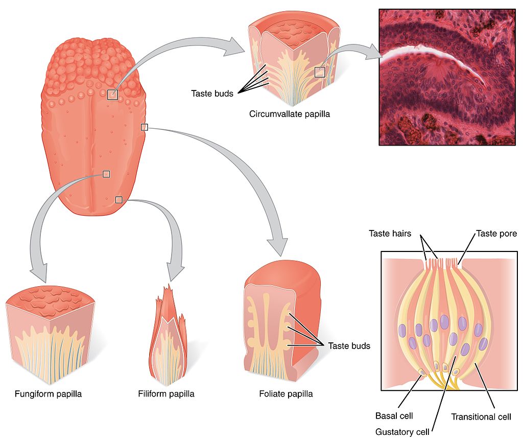
There are five basic tastes detected by the chemoreceptor cells in taste buds: saltiness, sourness, bitterness, sweetness, and umami (often described as a meaty taste). Contrary to popular belief, taste buds for the five basic tastes are not located on different parts of the tongue. Why does taste matter? The taste of food helps to stimulate the secretion of saliva from the salivary glands. It also helps us to eat foods that are good for us, instead of rotten or toxic foods. The detection of saltiness, for example, enables the control of salt intake and salt balance in the body. The detection of sourness may help us avoid spoiled foods, which often taste sour due to fermentation by bacteria. The detection of bitterness warns of poisons, because many plants defend themselves with toxins that taste bitter. The detection of sweetness guides us to foods that supply quick energy. The detection of umami may signal protein-rich foods.
Teeth
The teeth are complex structures made of a bone-like material called dentin and covered with enamel, which is the hardest tissue in the body. Adults normally have a total of 32 teeth, with 16 in each jaw. The right and left sides of each jaw are mirror images in terms of the numbers and types of teeth they contain. Teeth have different shapes to suit them for different aspects of mastication (chewing). The different types of teeth are illustrated in Figure 15.4.4.
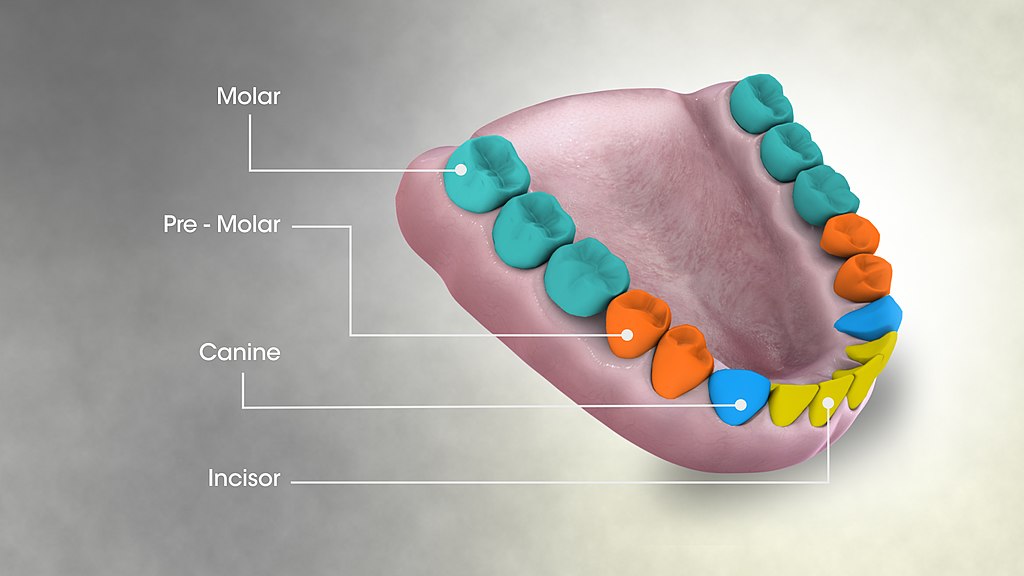
- Incisors are the sharp, blade-like teeth at the front of the mouth. They are used for cutting or biting off pieces of food. In adults, there are normally four incisors in each jaw, or eight in total.
- Canines are the pointed teeth on either side of the incisors. They are used for tearing foods that are tough or stringy. Adults normally have two canines in each jaw, or four altogether.
- Premolars and molars are cuboid teeth with cusps and grooves that are located on the sides and toward the back of the jaws. Premolars are closer to the front of the mouth. Molars are larger and have more cusps than premolars, but both are used for crushing and grinding food. Adults normally have two premolars and three molars on each side of each jaw, for a total of eight premolars and twelve molars.
Pharynx
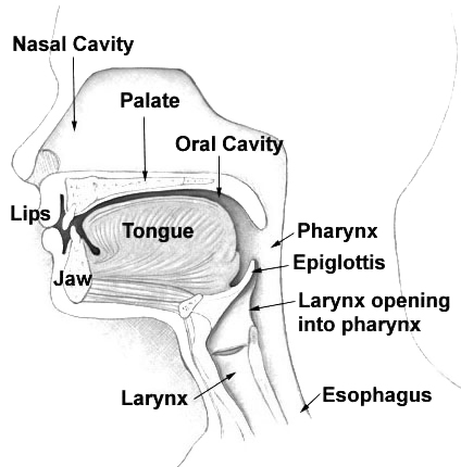
The tube-like pharynx (see Figure 15.4.5 below) plays a dual role as an organ of both respiration and digestion. As part of the respiratory system, it conducts air between the nasal cavity and larynx. As part of the digestive system, it allows swallowed food to pass from the oral cavity to the esophagus. Anything swallowed has priority over inhaled air when passing through the pharynx. During swallowing, the backward motion of the tongue causes a flap of elastic cartilage — called the epiglottis — to close over the opening to the larynx. This prevents food or drink from entering the larynx.
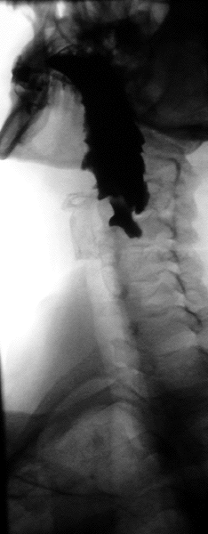
Esophagus
The esophagus (shown in Figure 15.4.6) is a muscular tube through which food is pushed from the pharynx to the stomach. The esophagus passes through an opening in the diaphragm (the large breathing muscle that separates the abdomen from the thorax) before reaching the stomach. In adults, the esophagus averages about 25 cm (about 9.8 inches) in length, depending on a person’s height. The inner lining of the esophagus consists of mucous membrane, which provides a smooth, slippery surface for the passage of food. The cells of this membrane are constantly being replaced as they are worn away from the frequent passage of food over them.
When food is not being swallowed, the esophagus is closed at both ends by upper and lower esophageal sphincters. Sphincters are rings of muscle that can contract to close off openings between structures. The upper esophageal sphincter is triggered to relax and open by the act of swallowing, allowing a bolus of food to enter the esophagus from the pharynx. Then, the esophageal sphincter closes again to prevent food from moving back into the pharynx from the esophagus.
Once in the esophagus, the food bolus travels down to the stomach, pushed along by the rhythmic contraction and relaxation of muscles (peristalsis). The lower esophageal sphincter is located at the junction between the esophagus and the stomach. This sphincter opens when the bolus reaches it, allowing the food to enter the stomach. The sphincter normally remains closed at other times to prevent the contents of the stomach from entering the esophagus. Failure of this sphincter to remain completely closed can lead to heartburn. If it happens chronically, it can lead to gastroesophageal reflux disease (GERD), in which the mucous membrane of the esophagus may become damaged by the highly acidic contents of the stomach.
See the video below to see how the parts of the upper GI tract work together to carry out swallowing:
Swallowing, uploaded by Alejandra Cork, 2012.
Stomach
The stomach is a J-shaped organ (shown in Figure 15.4.7) that is joined to the esophagus at its upper end, and to the first part of the small intestine (duodenum) at its lower end. When the stomach is empty of food, it normally has a volume of about 75 millilitres, but it can expand to hold up to about a litre of food. Waves of muscle contractions (peristalsis) passing through the muscular walls of the stomach cause the food inside to be mixed and churned. The wall of the stomach has an extra layer of muscle tissue not found in other organs of the GI tract that helps it squeeze and mix the food. These movements of the stomach wall contribute greatly to mechanical digestion by breaking the food into much smaller pieces. The churning also helps mix the food with stomach secretions that aid in its chemical digestion.
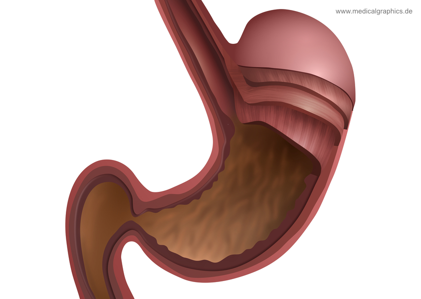
Secretions of the stomach include gastric acid, which consists mainly of hydrochloric acid (HCl). This makes the stomach contents highly acidic, which is necessary so that the enzyme pepsin — also secreted by the stomach — can begin the digestion of protein. Mucus is secreted by the lining of stomach to provide a slimy protective coating against the otherwise damaging effects of gastric acid. The fat-digesting enzyme lipase is secreted in small amounts in the stomach, but very little fat digestion occurs there.
By the time food has been in the stomach for about an hour, it has become the thick, semi-liquid chyme. When the small intestine is ready to receive chyme, a sphincter between the stomach and duodenum — called the pyloric sphincter — opens to allow the chyme to enter the small intestine for further digestion and absorption.
Feature: Reliable Sources
The ongoing epidemic of obesity in the wealthier nations of the world, including Canada, has led to the development of several different bariatric surgeries that modify the stomach to help obese patients reduce their food intake and lose weight. Go online to learn more about bariatric surgery. Find sources you judge to be reliable that answer the following questions:
- Who qualifies for bariatric surgery?
- Describe the bariatric surgeries commonly called stomach stapling, lap band, and gastric sleeve. How does each type of surgery modify the stomach? In terms of weight loss, how effective is each type?
- What are the major potential risks of bariatric surgery?
- Besides weight loss, what other benefits have been shown to result from bariatric surgery?
15.4 Summary
- Organs of the upper gastrointestinal (GI) tract include the mouth, pharynx, esophagus, and stomach.
- The mouth is the first organ of the GI tract. It has several structures that are specialized for digestion, including salivary glands, tongue, and teeth. Both mechanical digestion and chemical digestion of carbohydrates and fats begin in the mouth.
- The pharynx and esophagus move food from the mouth to the stomach, but are not involved in the process of digestion or absorption. Food moves through the esophagus by peristalsis.
- Mechanical and chemical digestion continue in the stomach. Acid and digestive enzymes secreted by the stomach start the chemical digestion of proteins. The stomach turns masticated food into a semi-fluid mixture called chyme.
15.4 Review Questions
-
- Identify structures in the mouth that are specialized for digestion.
- Describe digestion in the mouth.
- What general role do the pharynx and esophagus play in the digestion of food?
- How does food travel through the esophagus?
- Describe digestion in the stomach.
- Describe the differences between how air and food normally move past the pharynx.
- Name two structures in the mouth that contribute to mechanical digestion.
- What structure normally keeps stomach contents from backing up into the esophagus?
- Thirty minutes after you eat a meal, where is most of your food located? Explain your answer.
- What are two roles of mucus in the upper GI tract?
15.4 Explore More
What causes cavities? – Mel Rosenberg, TED-Ed, 2016.
How does alcohol make you drunk? – Judy Grisel, TED-Ed, 2020.
Gastric Bypass Surgery: One Patient’s Journey – Mayo Clinic, 2014.
Here’s What Happens In Your Body When You Swallow Gum | The Human Body, Tech Insider, 2018.
Attributions
Figure 15.4.1
Handstand, Pender Island, B.C. [photo] by Jasper Garratt on Unsplash is used under the Unsplash License (https://unsplash.com/license).
Figure 15.4.2
Blausen_0780_SalivaryGlands by BruceBlaus on Wikimedia Commons is used under a CC BY 3.0 (https://creativecommons.org/licenses/by/3.0) license.
Figure 15.4.3
1402_The_Tongue by OpenStax on Wikimedia Commons is used under a CC BY 4.0 (https://creativecommons.org/licenses/by/4.0) license.
Figure 15.4.4
1024px-3D_Medical_Animation_Still_Showing_Types_of_Teeth by http://www.scientificanimations.com on Wikimedia Commons is used under a CC BY-SA 4.0 (https://creativecommons.org/licenses/by-sa/4.0) license.
Figure 15.4.5
Illu01_head_neck by Arcadian from NCI/ SEER Training Modules on Wikimedia Common is in the public domain (https://en.wikipedia.org/wiki/public_domain).
Figure 15.4.6
ZenkerSchraeg by Bernd Brägelmann Braegel on Wikimedia Commons is used under a CC BY 3.0 (https://creativecommons.org/licenses/by/3.0) license. (Courtesy of Dr. Martin Steinhoff. It is not known whether there is a possibly necessary approval from the patient.)
Figure 15.4.7
Anatomy stomach – white by www.medicalgraphics.de from MedicalGraphics is used under a CC BY-ND 4.0 (https://creativecommons.org/licenses/by-nd/4.0/) license.
References
Alejandra Cork. (2012). Swallowing. YouTube. https://www.youtube.com/watch?v=pNcV6yAfq-g&t=4s
Betts, J. G., Young, K.A., Wise, J.A., Johnson, E., Poe, B., Kruse, D.H., Korol, O., Johnson, J.E., Womble, M., DeSaix, P. (2016, May 27). Figure 14.3 The tongue [digital image]. In Anatomy and Physiology (Section 14.1). OpenStax. https://openstax.org/books/anatomy-and-physiology/pages/14-1-sensory-perception
Blausen.com Staff. (2014). Medical gallery of Blausen Medical 2014. WikiJournal of Medicine 1 (2). DOI:10.15347/wjm/2014.010. ISSN 2002-4436.
Mayo Clinic. (2014, August 26). Gastric bypass surgery: One patient’s journey – Mayo Clinic. https://www.youtube.com/watch?v=twJBEypJDfU&feature=youtu.be
Mayo Clinic Staff. (n.d.). Gastroesophageal reflux disease (GERD) [online article]. MayoClinic.org. https://www.mayoclinic.org/diseases-conditions/gerd/symptoms-causes/syc-20361940
Tech Insider. (2018, March 20). Here’s what happens in your body when you swallow gum | The human body. YouTube. https://www.youtube.com/watch?v=u_1sVri3b2w&feature=youtu.be
TED-Ed. (2020, April 9). How does alcohol make you drunk? – Judy Grisel. YouTube. https://www.youtube.com/watch?v=gCrmFbgT37I&feature=youtu.be
TED-Ed. (2016, October 17). What causes cavities? – Mel Rosenberg. YouTube. https://www.youtube.com/watch?v=zGoBFU1q4g0&feature=youtu.be
A long, narrow, tube-like digestive organ through which food passes from the pharynx to the stomach.
A distinctive pattern of smooth muscle contractions that propels foodstuffs distally through the esophagus and intestines.
The part of the gastrointestinal tract that includes the mouth, pharynx, esophagus, and stomach.
The physical breakdown of chunks of food into smaller pieces by organs of the digestive system, for example chewing food.
Chemical breakdown of large, complex food molecules into smaller, simpler nutrient molecules that can be absorbed by blood or lymph. Usually involves a digestive enzyme.
The opening in the lower part of the human face, surrounded by the lips, through which food is taken in and from which speech and other sounds are emitted.
Epithelial tissue that lines inner body surfaces and body openings and produces mucus.
An involuntary, nonstriated muscle that is found in the walls of internal organs such as the stomach.
One of many exocrine glands in the mouth that secrete saliva into the mouth through ducts.
Gland such as a sweat gland, salivary gland, or mammary gland that secretes a substance into a duct that carries the secretion to the outside of the body.
A fluid secreted by salivary glands that keeps the mouth moist and contains the digestive enzymes amylase and lipase.
Either of a pair of large salivary glands situated just in front of each ear.
A salivary gland that is located deep under the mandible (jawbone).
A salivary gland that is located under the floor of the mouth, close to the midline.
The fleshy muscular organ in the mouth used for tasting, licking, swallowing, and articulating speech.
An enzyme, found chiefly in saliva and pancreatic fluid, that converts starch and glycogen into simple sugars.
A pancreatic enzyme that catalyzes the breakdown of fats to fatty acids and glycerol.
An antibody, also known as an immunoglobulin, is a large, Y-shaped protein produced mainly by plasma cells that is used by the immune system to neutralize pathogens such as pathogenic bacteria and viruses.
Nodules on the surface of the tongue that increase the surface area for the taste buds. Not all papillae, however, contain taste buds. The papillae also appear to aid in the mechanical handling of food, providing a rough surface.
A type of sensory receptor that responds to presence of chemicals.
A hard structure, embedded in the jaws of the mouth, that functions in chewing. Made of a dentin and covered in enamel, the hardest tissue in the body.
One of eight (four upper and four lower) blade-like teeth at the front of the mouth that are used to slice off pieces of food.
One of four pointed teeth on either side of the front teeth that are used for tearing foods.
One of eight cusped teeth in the sides of the jaws between the canine teeth and molars that are used for crushing food.
One of twelve teeth with cusps in the back of the mouth behind the premolars used for crushing and grinding food.
Tubular organ that connects the mouth and nasal cavity with the larynx and through which air and food pass.
The body system responsible for taking in oxygen and expelling carbon dioxide. The primary organs of the respiratory system are the lungs, which carry out this exchange of gases as we breathe.
A large, air-filled space in the skull above and behind the nose that helps conduct air in and out of the body as part of the upper respiratory tract.
An organ of the respiratory system between the pharynx and trachea, also called the voice box because it contains the vocal cords that allow the production of vocal sounds.
A body system including a series of hollow organs joined in a long, twisting tube from the mouth to the anus. The hollow organs that make up the GI tract are the mouth, esophagus, stomach, small intestine, large intestine, and anus. The liver, pancreas, and gallbladder are the solid organs of the digestive system.
A flap of cartilage at the root of the tongue, which is depressed during swallowing to cover the opening of the windpipe.
A large, dome-shaped muscle below the lungs that allows breathing to occur when it alternately contracts and relaxes.
A sac-like organ of the digestive system between the esophagus and small intestine in which both mechanical and chemical digestion take place.
A ring of muscles that can contract to close off an opening between structures, such as between the esophagus and stomach.
A lump of swallowed food.
A long, narrow, tube-like organ of the digestive system where most chemical digestion of food and virtually all absorption of nutrients take place.
The first and shortest of three parts of the small intestine where most chemical digestion occurs.
The chief digestive enzyme in the stomach, which breaks down proteins into polypeptides.
A class of biological molecule consisting of linked monomers of amino acids and which are the most versatile macromolecules in living systems and serve crucial functions in essentially all biological processes.
A slimy substance produced by mucous membranes that traps pathogens, particles, and debris.
A thick, semi-liquid mixture that food in the gastrointestinal tract becomes by the time it leaves the stomach.
