11.6 Joints
Created by CK-12 Foundation/Adapted by Christine Miller
Double Jointed?
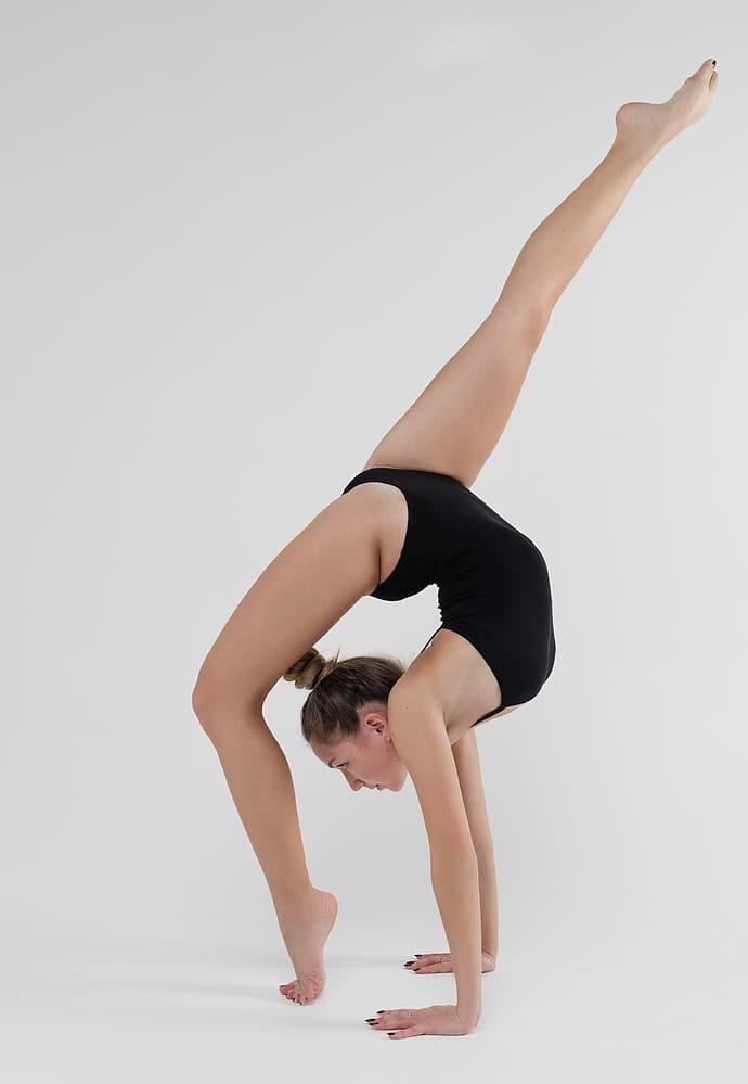
Is this woman double jointed? No, there is actually no such thing — at least as far as humans are concerned. However, some people, like the woman pictured in Figure 11.6.1, are much more flexible than others, generally because they have looser ligaments. Physicians call this condition joint hypermobility. Regardless of what it’s called, the feats of people with highly mobile joints can be quite impressive.
What Are Joints?
Joints are locations at which bones of the skeleton connect with one another. A joint is also called an articulation. The majority of joints are structured in such a way that they allow movement. However, not all joints allow movement. Of joints that do allow movement, the extent and direction of the movements they allow also vary.
Classification of Joints
Joints can be classified structurally or functionally. The structural classification of joints depends on the manner in which the bones connect to each other. The functional classification of joints depends on the nature of the movement the joints allow. There is significant overlap between the two types of classifications, because function depends largely on structure.
Structural Classification of Joints
The structural classification of joints is based on the type of tissue that binds the bones to each other at the joint. There are three types of joints in the structural classification: fibrous, cartilaginous, and synovial joints.
- Fibrous joints are joints in which bones are joined by dense connective tissue that is rich in collagen fibres. These joints are also called sutures. The joints between bones of the cranium are fibrous joints.
- Cartilaginous joints are joints in which bones are joined by cartilage. The joints between most of the vertebrae in the spine are cartilaginous joints.
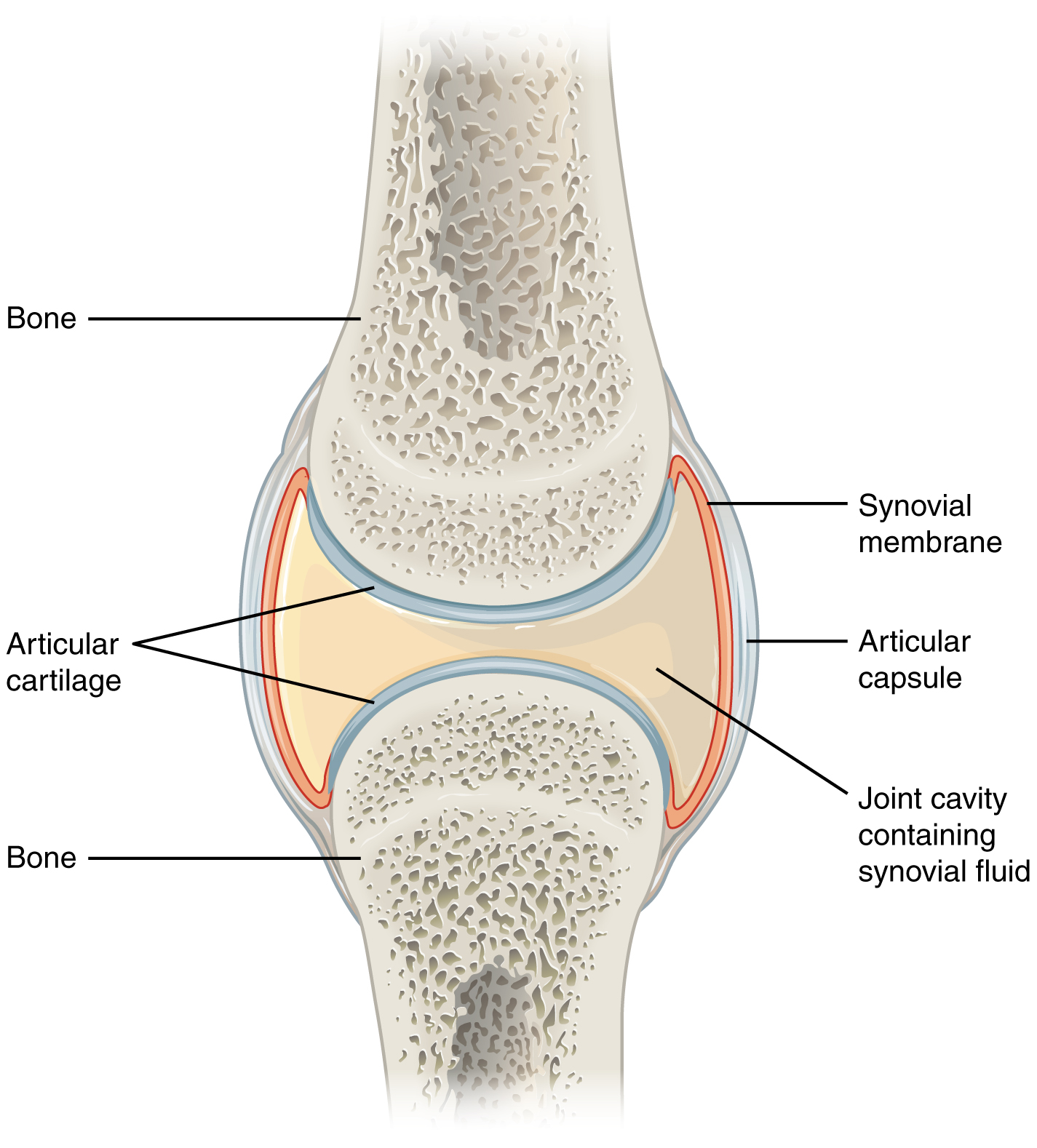
- Synovial joints are characterized by a fluid-filled space (called a synovial cavity) between the bones of the joints. You can see a drawing of a typical synovial joint in Figure 11.6.2. The cavity is enclosed by a membrane and filled with a fluid (called synovial fluid) that provides extra cushioning to the ends of the bones. Cartilage covers the articulating surfaces of the two bones, but the bones are actually held together by ligaments. The knee is a synovial joint.
Functional Classification of Joints
The functional classification of joints is based on the type and degree of movement that they allow. There are three types of joints in the functional classification: immovable, partly movable, and movable joints.
- Immovable joints allow little or no movement at the joint. Most immovable joints are fibrous joints. Besides the bones of the cranium, immovable joints include joints between the tibia and fibula in the lower leg, and between the radius and ulna in the lower arm.
- Partly movable joints permit slight movement. Most partly movable joints are cartilaginous joints. Besides the joints between vertebrae, they include the joints between the ribs and sternum (breast bone).
- Movable joints allow bones to move freely. All movable joints are synovial joints. Besides the knee, they include the shoulder, hip, and elbow. Movable joints are the most common type of joints in the body.
Types of Movable Joints
Movable joints can be classified further according to the type of movement they allow. There are six classes of movable joints: pivot, hinge, saddle, plane, condyloid, and ball-and-socket joints. An example of each class — as well as the type of movement it allows — is shown in Figure 11.6.3.
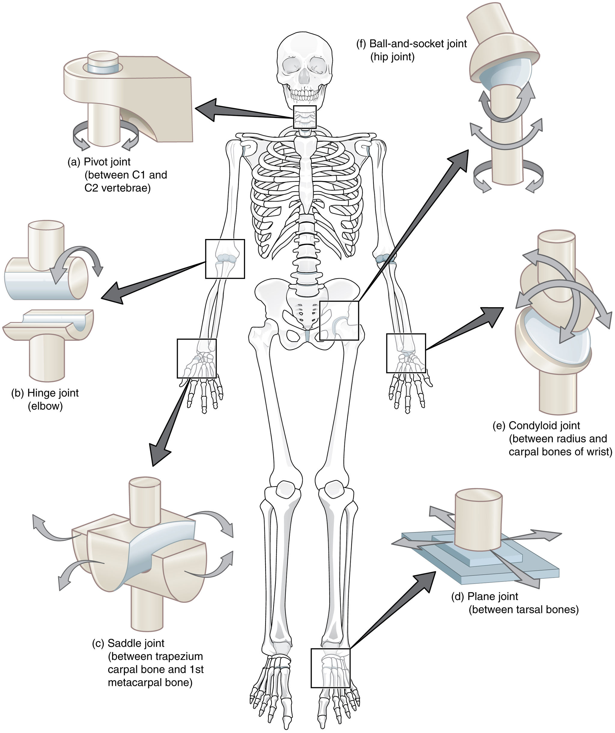
- A ball-and-socket joint allows the greatest range of movement of any movable joint. It allows forward and backward motion, as well as upward and downward movement. It also allows rotation in a circle. The hip and shoulder are the only two ball-and-socket joints in the human body.
- A pivot joint allows one bone to rotate around another. An example of a pivot joint is the joint between the first two vertebrae in the spine. This joint allows the head to rotate from left to right and back again.
- A hinge joint allows back and forth movement like the hinge of a door. An example of a hinge joint is the elbow. This joint allows the arm to bend back and forth.
- A saddle joint allows two different types of movement. An example of a saddle joint is the joint between the first metacarpal bone in the hand and one of the carpal bones in the wrist. This joint allows the thumb to move toward and away from the index finger, and also to cross over the palm toward the little finger.
- A plane joint (also called a gliding joint) allows two bones to glide over one another. The joints between the tarsals in the ankles and between the carpals in the wrists are mainly gliding joints. In the wrist, this type of joint allows the hand to bend upward at the wrist, and also to wave from side to side while the lower arm is held steady.
- A condyloid joint is one in which an oval-shaped head on one bone moves in an elliptical cavity in another bone, allowing movement in all directions, except rotation around an axis. The joint between the radius in the lower arm and carpal bones of the wrist is a condyloid joint, as is the joint at the base of the index finger.
Feature: My Human Body
Of all the parts of the skeletal system, the joints are generally the most fragile and subject to damage. If the cartilage that cushions bones at joints wears away, it does not grow back. Eventually, all of the cartilage may wear away. This causes osteoarthritis, which can be both painful and debilitating. In serious cases of osteoarthritis, people may lose the ability to climb stairs, walk long distances, perform routine daily activities, or participate in activities they love, such as gardening or playing sports. If you protect your joints, you can reduce your chances of joint damage, pain, and disability. If you already have joint damage, it is equally important to protect your joints and limit further damage. Follow these five tips:
- Maintain a normal, healthy weight. The more you weigh, the more force you exert on your joints. When you walk, each knee has to bear a force equal to as much as six times your body weight. If a person weighs 200 pounds, each knee bears more than half a ton of weight with every step. Seven in ten knee replacement surgeries for osteoarthritis can be attributed to obesity.
- Avoid too much high-impact exercise. Examples of high-impact activities include volleyball, basketball, and tennis. These activities generally involve running or jumping on hard surfaces, which puts tremendous stress on weight-bearing joints, especially the knees. Replace some or all of your high-impact activities with low-impact activities, such as biking, swimming, yoga, or lifting light weights.
- Reduce your risk of injury. Don’t be a weekend warrior, sitting at a desk all week and then crowding all your physical activity into two days. Get involved in a regular, daily exercise routine that keeps your body fit and your muscles toned. Building up muscles will make your joints more stable, allowing stress to spread across them. Be sure to do some stretching every day to keep the muscles around joints flexible and less prone to injury.
- Distribute work over your body, and use your largest, strongest joints. Use your shoulder, elbow, and wrist to lift heavy objects — not just your fingers. Hold small items in the palm of your hand, rather than by the fingers. Carry heavy items in a backpack, rather than in your hands. Hold weighty objects close to your body, instead of at arms’ length. Lift with your hips and knees, not your back.
- Respect pain. If it hurts, stop doing it. Take a break from the activity — at least until the pain stops. Try to use joints only to the point of mild fatigue, not pain.
11.6 Summary
- Joints are spots at which bones of the skeleton connect with one another. A joint is also called an articulation.
- Joints can be classified structurally or functionally, and there is significant overlap between the two types of classifications.
- The structural classification of joints depends on the type of tissue that binds the bones to each other at the joint. There are three types of joints in the structural classification: fibrous, cartilaginous, and synovial joints.
- The functional classification of joints is based on the type and degree of movement that they allow. There are three types of joints in the functional classification: immovable, partly movable, and movable joints.
- Movable joints can be classified further according to the type of movement they allow. There are six classes of movable joints: pivot, hinge, saddle, plane, condyloid, and ball-and-socket joints.
11.6 Review Questions
- What are joints?
- What are two ways that joints are commonly classified?
-
- How are joints classified structurally?
- Describe the functional classification of joints.
- How are movable joints classified?
- Name the six classes of movable joints. Describe how they move and give an example of each.
- Which specific type of moveable joint do you think your knee joint is? Explain your reasoning.
- Explain the difference between cartilage in a cartilaginous joint and cartilage in a synovial joint.
- Why are fibrous joints immovable?
- What is the function of synovial fluid?
11.6 Explore More
Why do your knuckles pop? – Eleanor Nelsen, TED-Ed, 2015.
Why haven’t we cured arthritis? – Kaitlyn Sadtler and Heather J. Faust, TED-Ed, 2019.
Attributions
Figure 11.6.1
Tags: Sports Gymnastics Fitness Woman Preparation by nastya_gepp on Pixabay is used under the Pixabay License (https://pixabay.com/de/service/license/).
Figure 11.6.2
Synovial_Joints by OpenStax College on Wikimedia Commons is used under a CC BY 3.0 (https://creativecommons.org/licenses/by/3.0) license.
Figure 11.6.3
Types_of_Synovial_Joints by OpenStax College on Wikimedia Commons is used under a CC BY 3.0 (https://creativecommons.org/licenses/by/3.0) license.
References
Betts, J. G., Young, K.A., Wise, J.A., Johnson, E., Poe, B., Kruse, D.H., Korol, O., Johnson, J.E., Womble, M., DeSaix, P. (2013, June 19). Figure 9.8 Synovial joints [digital image]. In Anatomy and Physiology (Section 9.4). OpenStax. https://openstax.org/books/anatomy-and-physiology/pages/9-4-synovial-joints
Betts, J. G., Young, K.A., Wise, J.A., Johnson, E., Poe, B., Kruse, D.H., Korol, O., Johnson, J.E., Womble, M., DeSaix, P. (2013, June 19). Figure 9.10 Types of synovial joints [digital image]. In Anatomy and Physiology (Section 9.4). OpenStax. https://openstax.org/books/anatomy-and-physiology/pages/9-4-synovial-joints
Mayo Clinic Staff. (n.d.). Osteoarthritis [online article]. MayoClinic.org. https://www.mayoclinic.org/diseases-conditions/osteoarthritis/symptoms-causes/syc-20351925
TED-Ed. (2015, May 5). Why do your knuckles pop? – Eleanor Nelsen. YouTube. https://www.youtube.com/watch?v=IjiKUmfaZr4
TED-Ed. (2019, November 7). Why haven’t we cured arthritis? – Kaitlyn Sadtler and Heather J. Faust. YouTube. https://www.youtube.com/watch?v=FWsBm3hr3B0
A structure where two or more bones of the skeleton come together.
An immovable joint in which bones are connected by collagen fibers; also called a suture.
A partly movable joint in which bones are joined by cartilage.
A movable joint in which a fluid-filled synovial cavity separates bones at the joint.
An articulation between bones in which no movement occurs. It is also referred to as synarthrotic (meaning immovable).
An articulation between bones (also called an amphiarthrotic joint) in which the motion is limited due to either fibrous tissue or cartilage.
A joint in which the opposing bony surfaces are covered with a layer of hyaline cartilage or fibrocartilage and in which some degree of free movement is possible.
A natural or manufactured joint or coupling, such as the hip joint, in which a partially spherical end lies in a socket, allowing multi-directional movement and rotation.
The joints that allow bones to rotate. In a pivot joint, a cylinder shaped bone rotates inside another bone or ligament that forms a ring around it.
A synovial bone joint in which the articular surfaces are molded to each other in such a manner as to permit motion only in one plane.
A type of synovial joint that allow articulation by reciprocal reception. Both bones have concave-convex articular surfaces which interlock like two saddles opposed to one another (example, the base of the thumb).
A type of structure in the body, also called gliding joint, formed between two bones in which the articular, or free, surfaces of the bones are flat or nearly flat, enabling the bones to slide over each other.
A synovial joint in which an oval-shaped process of one bone fits into a roughly elliptical cavity of the other, allowing movement in two planes.
The degeneration of joint cartilage and the underlying bone, most common from middle age onward. It causes pain and stiffness, especially in the hip, knee, and thumb joints.
A rigid organ that constitutes part of the vertebrate skeleton in animals.
