11.3 Divisions of the Skeletal System
Created by CK-12 Foundation/Adapted by Christine Miller
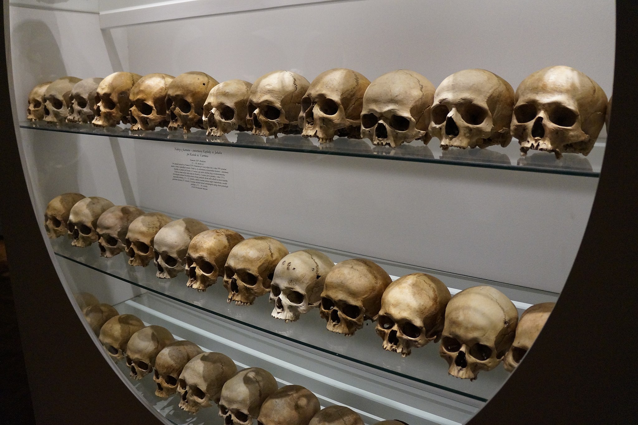
Skulls on Display
This somewhat macabre display (Figure 11.3.1) can be viewed at the Slovak National Museum in Bratislava, Slovakia. The skulls are meant to represent normal human skeletal anatomy. The skull is part of the axial skeleton, which is one of the two major divisions of the human skeleton. The other division is the appendicular skeleton.
Axial Skeleton
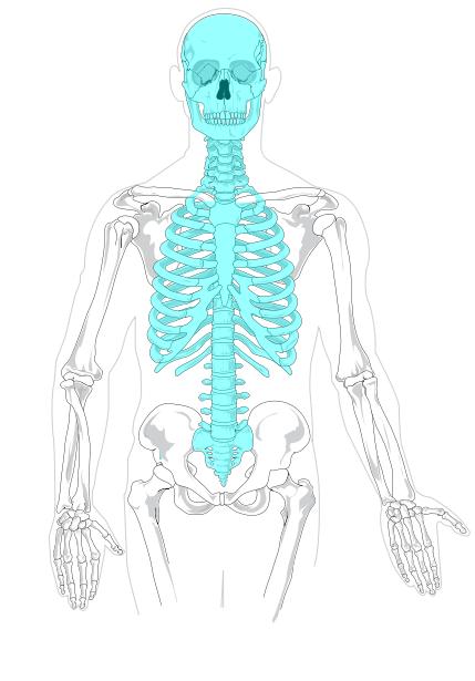
The axial skeleton, shown in blue in Figure 11.3.2, consists of a total of 80 bones. Besides the skull, it includes the rib cage and vertebral column. It also includes the three tiny ossicles (hammer, anvil, and stirrup) in the middle ear and the hyoid bone in the throat, to which the tongue and some other soft tissues are attached.
Skull
The skull is the part of the human skeleton that provides a bony framework for the head. It consists of 22 different bones. There are eight bones in the cranium, which encloses the brain, and 14 bones in the face.
Cranium
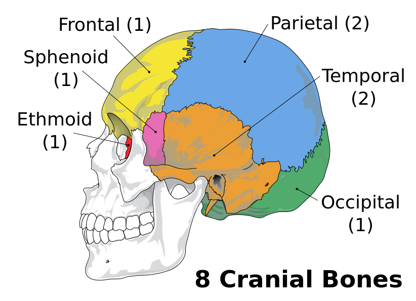
The cranium forms the entire upper portion of the skull. As shown in Figure 11.3.3, it consists of eight bones: one frontal bone, two parietal bones, two temporal bones, one occipital bone, one sphenoid bone, and one ethmoid bone. The ethmoid bone separates the nasal cavity from the brain. The sphenoid bone is one of several bones, including the frontal bone, that help form the eye sockets. The other bones of the cranium are large and plate-like. They cover and protect the brain. The bottom of the skull has openings for major blood vessels and nerves. A large opening, called the foramen, connects the spinal cord and brain.
Facial Bones
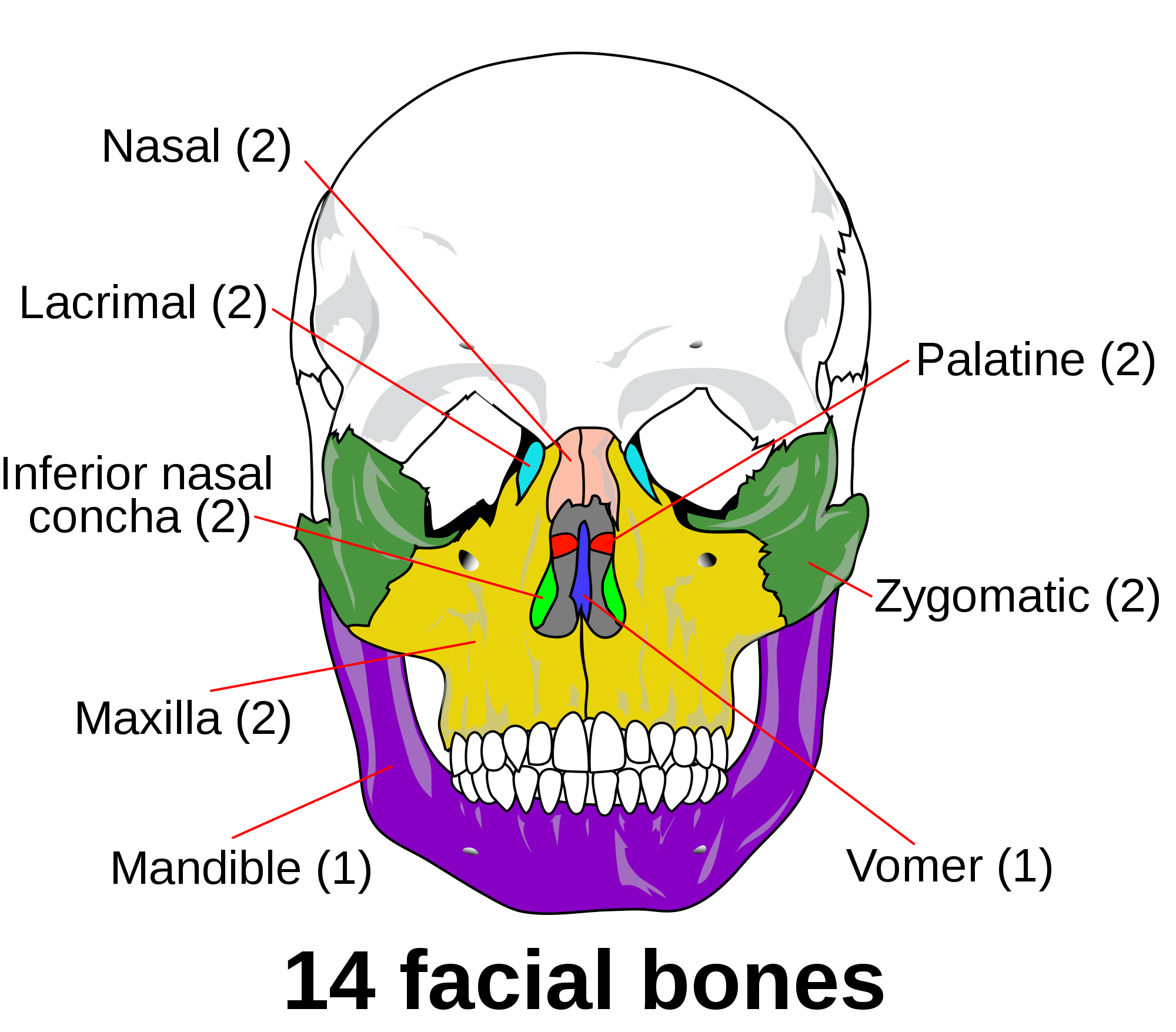
The 14 facial bones of the skull are located below the frontal bone of the cranium, and they are depicted in Figure 11.3.4. Large bones in the face include the upper jaw bones, or maxillae (singular, maxilla), which form the middle part of the face and the bottom of the two eye sockets. The maxillae are fused together, except for an opening between them for the nose. The lower edge of the maxillae contains sockets for the upper teeth. The lower jaw bone, or mandible, is also large. The top edge of the mandible contains sockets for the lower teeth. The mandible opens and closes to chew food and is controlled by strong muscles. There are two zygomatic (or cheek) bones and two nasal bones. The nasal region also contains seven smaller bones, as indicated in Figure 11.3.4.
Vertebral Column
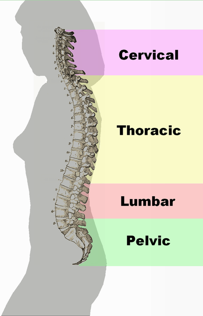
The vertebral column — also called the spine or backbone — is the flexible column of vertebrae (singular, vertebra) that connects the trunk with the skull and encloses the spinal cord. It consists of 33 vertebrae that are divided into five regions, as shown in Figure 11.3.5: the cervical, thoracic, lumbar, sacral, and coccygeal regions. From the neck down, the first 24 vertebrae (cervical, thoracic, and lumbar) are individual bones. The five sacral vertebrae are fused together, as are the four coccygeal vertebrae.
The vertebral column consists of 24 individual vertebrae that are separated by intervertebral discs of cartilage. An additional nine vertebrae are fused together at the base of the spine. Note the S-shaped curve of the vertebral column in the profile view in Figure 11.3.5 on the left.
The human vertebral column reflects adaptations for upright bipedal locomotion (walking upright on two legs). For example, the vertebral column is less like a rigid column than an S-shaped spring (see profile view in Figure 11.3.5). Although newborn infants have a relatively straight spine, the curves develop as the backbone starts taking on its support functions, such as keeping the trunk erect, holding up the head, and helping to anchor the limbs. The S shape of the vertebral column allows it to act like a shock absorber, absorbing much of the jarring of walking and running so the forces are not transmitted directly from the pelvis to the skull. The S shape also helps protect the spine from breaking, which would be more likely with a straight, more rigid vertebral column. In addition, the S shape helps to distribute the weight of the body — particularly of the internal organs, so the weight load is not all at the bottom, as would occur with a straight spine.
Rib Cage
The rib cage (also called thoracic cage) is aptly named, because it forms a sort of cage that holds within it the organs of the upper part of the trunk, including the heart and lungs. It is shown in Figures 11.3.6–11.3.8. The rib cage includes the 12 thoracic vertebrae and the sternum, as well as 12 pairs of ribs, which are attached at joints to the vertebrae. The ribs are divided into three groups, called true ribs, false ribs, and floating ribs. The top seven pairs of ribs are true ribs. They are attached by cartilage directly to the sternum. The next three pairs of ribs are false ribs. They are attached by cartilage to the ribs above them, rather than directly to the sternum. The lowest two pairs of ribs are floating ribs. They are attached by cartilage to muscles in the abdominal wall. The attachments of false and floating ribs let the lower part of the rib cage expand to accommodate the internal movements of breathing.
 |
 |
 |
Appendicular Skeleton
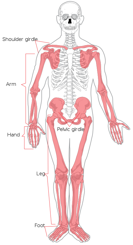
The appendicular skeleton, shown in red (Figure 11.3.9), consists of a total of 126 bones. It includes all the bones of the limbs (arms, legs, hands, and feet,) as well as the bones of the shoulder (shoulder girdle) and pelvis (pelvic girdle).
Upper Limbs
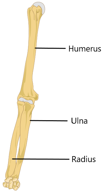
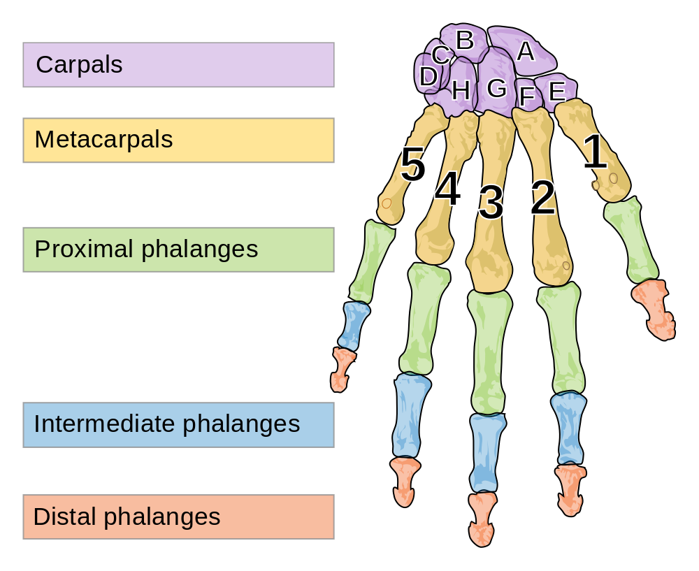
Each upper limb consists of 30 bones. As shown in Figure 11.3.10, there is one bone (called the humerus) in each of the upper arms, and there are two bones (called the ulna and radius) in each of the lower arms. The remaining bones of the upper limb are shown in Figure 11.3.11. Each wrist contains eight carpal bones — which are arranged in two rows of four bones each — and each hand contains five metacarpal bones. The bones in the fingers of each hand include 14 phalanges (three in each finger except the thumb, which has two phalanges). The thumb has the unique ability to move into opposition with the palm of the hand, and with each of the fingers when they are slightly bent. This allows the hand to handle and manipulate objects such as tools.
Lower Limbs
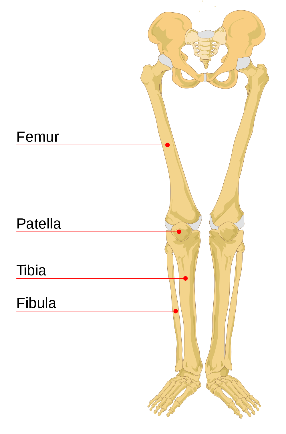
Each lower limb consists of 30 bones. As shown in Figure 11.3.12 to the left, there is one bone (called the femur) in each of the upper legs, and there are two bones (called the tibia and fibula) in each of the lower legs. The knee cap (or patella) is an additional leg bone at the front of each knee, which is the largest joint in the human body.
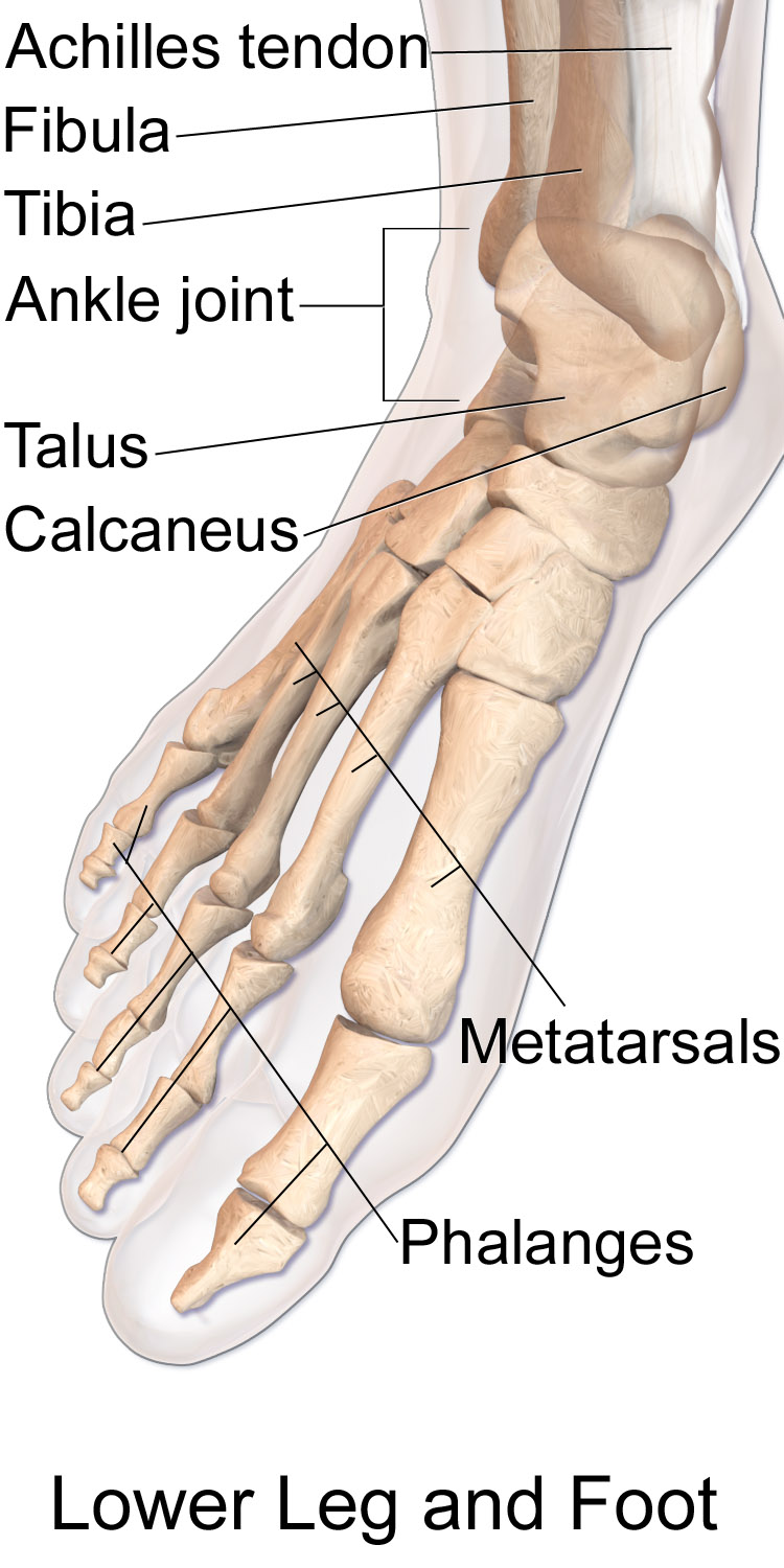
The remaining bones of the lower limbs are in Figure 11.3.13 to the right. Each ankle contains seven tarsal bones (including the talus and calcaneus), and each foot contains five metatarsal bones. The tarsals and metatarsals form the ankle, heel, and arch of the foot. They give the foot strength while allowing flexibility. The bones in the toes of each foot consist of 14 phalanges (three in each toe except the big toe, which has two phalanges)
Bones of the lower leg (fibula and tibia), ankle (talus), heel (calcaneus), foot (metatarsals), and toes (phalanges)
Shoulder Girdle
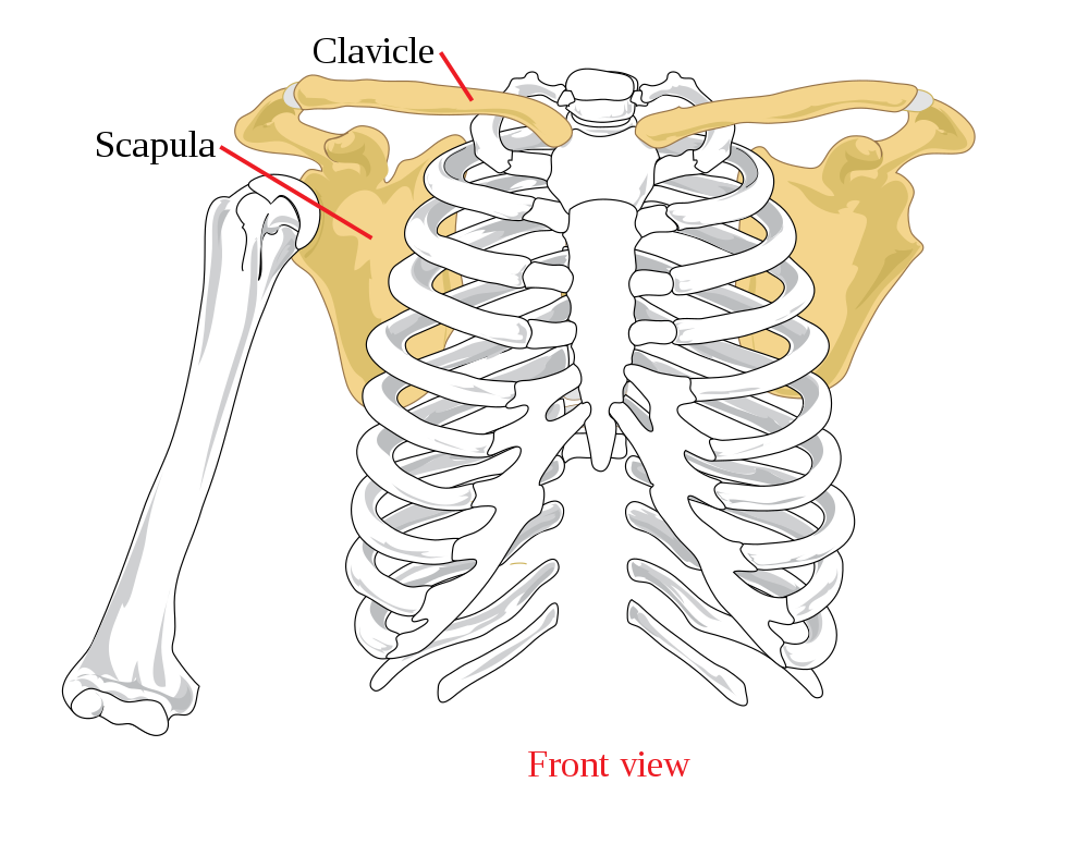
The pectoral girdle (also called shoulder girdle) attaches the upper limbs to the trunk of the body. It is connected to the axial skeleton by muscles alone. This allows a considerable range of motion in the upper limbs. The shoulder girdle consists of just two pairs of bones, with one of each pair on opposite sides of the body (see Figure 11.3.14). There are a right and left clavicle (collarbone), and a right and left scapula (shoulder blade). The scapula is a pear-shaped flat bone that helps form the shoulder joint. The clavicle is a long bone that serves as a strut between the shoulder blade and the sternum.
Pelvic Girdle
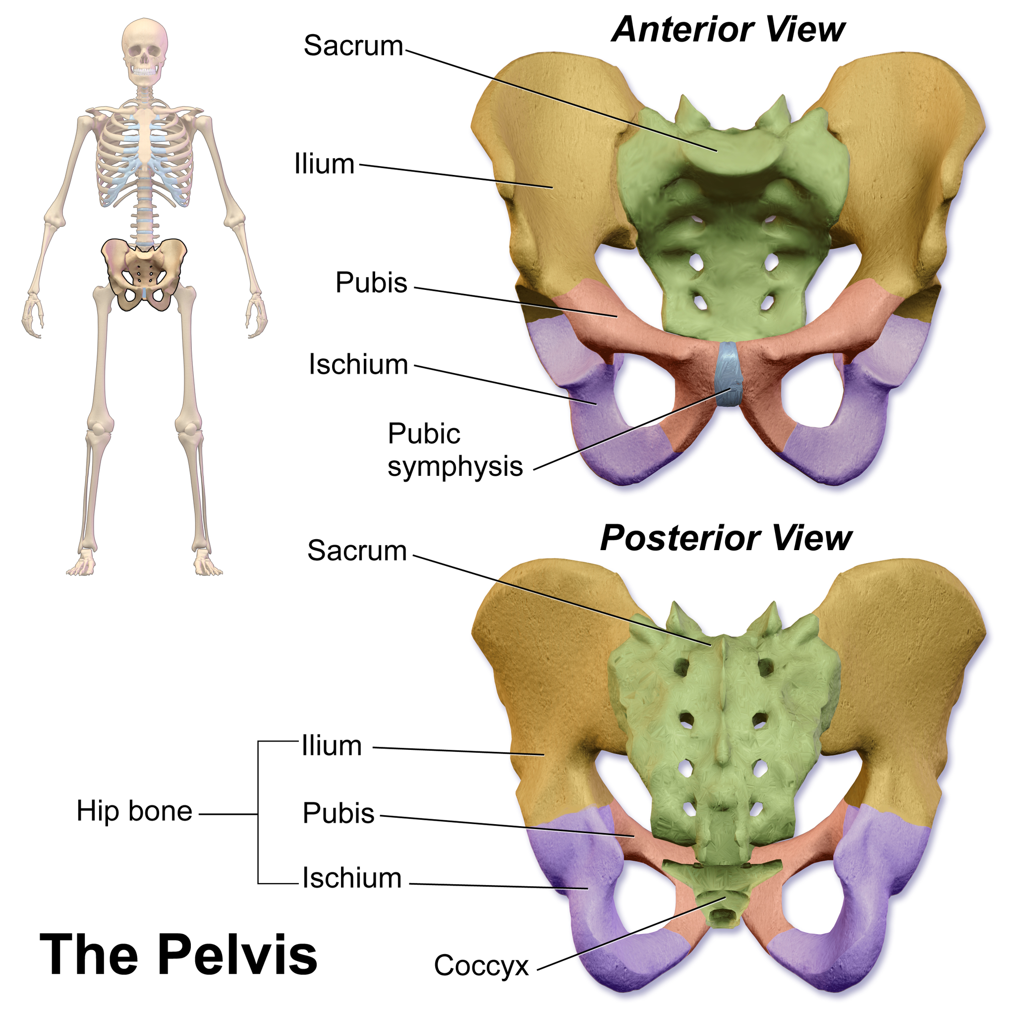
The pelvic girdle attaches the legs to the trunk of the body, and also provides a basin to contain and support the organs of the abdomen. It is connected to the vertebral column of the axial skeleton by ligaments. The pelvic girdle consists of two halves — one half for each leg — but the halves are fused with each other in adults at a joint called the pubic symphysis. Each half of the pelvic girdle includes three bones, as shown in Figure 11.3.15 to the right: the ilium (flaring upper part of the pelvic girdle), pubis (lower front), and ischium (lower back). Each of these bones helps form the acetabulum, which is a depression into which the top of the femur (thighbone) fits. When the body is in a seated position, it rests on protrusions (called tuberosities) of the two ischial bones.
11.3 Summary
- The axial skeleton consists of a total of 80 bones. It includes the skull, vertebral column, and rib cage. It also includes the three tiny ossicles in the middle ear and the hyoid bone in the throat.
- The skull provides a bony framework for the head. It consists of 22 different bones: eight in the cranium (which encloses the brain) and 14 in the face (which includes the upper and lower jaw).
- The vertebral column is a flexible, S-shaped column of 33 vertebrae that connects the trunk with the skull and encloses the spinal cord. The vertebrae are divided into five regions: cervical, thoracic, lumbar, sacral, and coccygeal regions. The S shape of the vertebral column allows it to absorb shocks and distribute the weight of the body.
- The rib cage holds and protects the organs of the upper part of the trunk, including the heart and lungs. It includes the 12 thoracic vertebrae, the sternum, and 12 pairs of ribs.
- The appendicular skeleton consists of a total of 126 bones. It includes the bones of the four limbs, shoulder girdle, and pelvic girdle.
- Each upper limb consists of 30 bones. There is one bone (called the humerus) in the upper arm, and two bones (called the ulna and radius) in the lower arm. The wrist contains eight carpal bones, the hand contains five metacarpals, and the fingers consist of 14 phalanges. The thumb is opposable to the palm and fingers of the same hand.
- Each lower limb also consists of 30 bones. There is one bone (called the femur) in the upper leg, and two bones (called the tibia and fibula) in the lower leg. The patella covers the knee joint. The ankle contains seven tarsal bones, and the foot contains five metatarsals. The tarsals and metatarsals form the heel and arch of the foot. The bones in the toes consist of 14 phalanges.
- The shoulder girdle attaches the upper limbs to the trunk of the body. It is connected to the axial skeleton only by muscles, allowing mobility of the upper limbs. Bones of the shoulder girdle include a right and left clavicle, as well as a right and left scapula.
- The pelvic girdle attaches the legs to the trunk of the body, and supports the organs of the abdomen. It is connected to the axial skeleton by ligaments. The pelvic girdle consists of two halves that are fused together in adults. Each half consists of three bones: the ilium, pubis, and ischium.
11.3 Review Questions
-
-
- What are the advantages of an S-shaped vertebral column?
- What is the rib cage? What is its function? What types of ribs are there?
- Explain the advantage of having some ribs that are not attached directly to the sternum.
- What is the shoulder girdle? Why does it allow considerable upper limb mobility?
- Describe some of the similarities between the upper limbs and the lower limbs.
- Describe the pelvic girdle and the bones it contains.
11.3 Explore More
Bones of the skull – Learn in 4 minutes! Neural Academy, 2018.
Craniosynostosis – Mayo Clinic, 2011.
Attributions
Figure 11.3.1
Human_skulls_on_display by Kiwiev on Wikimedia Commons is used under a CC0 1.0 Universal Public Domain Dedication (https://creativecommons.org/publicdomain/zero/1.0/deed.en) license.
Figure 11.3.2
Axial_skeleton_diagram_blank.svg by Quico/ Qllach on Wikimedia Commons is released into the public domain (https://en.wikipedia.org/wiki/Public_domain). (This is a derivative work from Axial skeleton diagram.svg, by Mariana Ruiz Villarreal [LadyofHats].)
Figure 11.3.3
822px-Cranial_bones_en_v2.svg by Was a bee (adapted/ reallocated text on original image File:Cranial bones en.svg. by Edoarado) on Wikimedia Commons is used under a CC0 1.0 Universal Public Domain Dedication (https://creativecommons.org/publicdomain/zero/1.0/deed.en) license.
Figure 11.3.4
Facial_skeleton_-_en.svg by Was a bee (adapted original image File:Es-Human skull front simplified (bones).svg. by Cristobal carrasco) on Wikimedia Commons is released into the public domain (https://en.wikipedia.org/wiki/Public_domain).
Figure 11.3.5
Spinal_column_curvature by vsion on Wikimedia Commons is in the public domain (https://en.wikipedia.org/wiki/Public_domain).
Figure 11.3.6
True_ribs_animation from en:Anatomography on Wikimedia Commons is used under a CC BY-SA 2.1 JP (https://creativecommons.org/licenses/by-sa/2.1/jp/deed.en) license. (Creator/ licensor: “BodyParts3D, © The Database Center for Life Science licensed under CC Attribution-Share Alike 2.1 Japan.”)
Figure 11.3.7
False_ribs_animation from en:Anatomography on Wikimedia Commons is used under a CC BY-SA 2.1 JP (https://creativecommons.org/licenses/by-sa/2.1/jp/deed.en) license. (Creator/ licensor: “BodyParts3D, © The Database Center for Life Science licensed under CC Attribution-Share Alike 2.1 Japan.”)
Figure 11.3.8
Floating_ribs_animation from en:Anatomography on Wikimedia Commons is used under a CC BY-SA 2.1 JP (https://creativecommons.org/licenses/by-sa/2.1/jp/deed.en) license. (Creator/ licensor: “BodyParts3D, © The Database Center for Life Science licensed under CC Attribution-Share Alike 2.1 Japan.”)
Figure 11.3.9
Appendicular_skeleton_diagram.svg by Mariana Ruiz Villarreal [LadyofHats] on Wikimedia Commons is released into the public domain (https://en.wikipedia.org/wiki/Public_domain).
Figure 11.3.10
Humerus,_ulna_and_radius_(female) by Mikael Häggström on Wikimedia Commons is used and adapted by Christine Miller (addition of labels), as it has been released into the public domain (https://en.wikipedia.org/wiki/Public_domain).
Figure 11.3.11
Human_left_hand_bones_with_metacarpal_numbers_and_carpal_letters.svg by Mariana Ruiz Villarreal [LadyofHats], Nyks, Bibi Saint-Pol. Bloubéri. and Whidou on Wikimedia Commons is used under a CC0 1.0 Universal Public Domain Dedication (https://creativecommons.org/publicdomain/zero/1.0/deed.en) license.
Figure 11.3.12
Human_leg_bones_labeled.svg by Jecowa (original uploader) at English Wikipediaon Wikimedia Commons is in the public domain (https://en.wikipedia.org/wiki/Public_domain).
Figure 11.3.13
Blausen_0411_FootAnatomy by BruceBlaus on Wikimedia Commons is used under a CC BY 3.0 (https://creativecommons.org/licenses/by/3.0) license.
Figure 11.3.14
Pectoral_girdle_front_diagram.svg by Mariana Ruiz Villarreal [LadyofHats] on Wikimedia Commons is released into the public domain (https://en.wikipedia.org/wiki/Public_domain).
Figure 11.3.15
2048px-Blausen_0723_Pelvis by BruceBlaus on Wikimedia Commons is used under a CC BY 3.0 (https://creativecommons.org/licenses/by/3.0) license.
References
Blausen.com staff. (2014). Medical gallery of Blausen Medical 2014. WikiJournal of Medicine 1 (2). DOI:10.15347/wjm/2014.010. ISSN 2002-4436.
Mayo Clinic. (2011, ). Craniosynostosis – Mayo Clinic. YouTube. https://www.youtube.com/watch?v=fcHB2pvH2uc
Neural Academy. (2018, ). Bones of the skull – Learn in 4 minutes! YouTube. https://www.youtube.com/watch?v=WRmNC_yPQZ8
The part of the human skeleton that provides a bony framework for the head and includes bones of the cranium and face.
A division of the skeleton that includes the skull, rib cage, and vertebral column.
The bones of the upper and lower limbs, shoulder girdle, and pelvic girdle.
The upper part of the skull that encloses and protects the brain.
A flexible column of vertebrae that connects the trunk to the skull and encloses the spinal cord; also called spine or backbone.
One of 33 small bones that make up the vertebral column.
The region of the spinal column containing the vertebrae of the neck, immediately below the skull.
each of the twelve bones of the backbone to which the ribs are attached.
Any of the five vertebrae situated between the thoracic vertebrae above and the sacrum below.
A large, triangular bone at the base of the spine that forms by the fusing of sacral vertebrae S1–S5 between 18 and 30 years of age.
Relating to the coccyx. The coccyx, also known as the tailbone, is a small, triangular bone resembling a shortened tail located at the bottom of the spine.The vertebrae may be fused together to form a single bone; however, in some cases, the first vertebra is separate from the others.
Discs which lie between adjacent vertebrae in the vertebral column. Each disc forms a fibrocartilaginous joint (a symphysis), to allow slight movement of the vertebrae, to act as a ligament to hold the vertebrae together, and to function as a shock absorber for the spine.
A bony “cage” enclosing the thoracic cavity and consisting of the ribs, thoracic vertebrae, and sternum.
A muscular organ in the chest that pumps blood through blood vessels when it contracts.
Two paired organs of the respiratory system in which gas exchange takes place between the blood and the atmosphere.
Paired clavicles (collar bones) and scapulas (shoulder blades) that together form the shoulders and attach the arms to the trunk; also called shoulder girdle.
Paired, fused bones (ilium, pubis, and ischium) that form the hips and attach the legs to the trunk.

.svg)
.png)