8.7 Human Senses
Created by CK-12 Foundation/Adapted by Christine Miller

Seeing Is Believing
At first glance, Figure 8.7.1 appears to be just random dots of colour, but hidden within it is the three-dimensional shape of a bee. Can you see it among the dots? This figure is an example of a stereogram, which is a two-dimensional picture that, when viewed correctly, reveals a three-dimensional object. If you can’t see the hidden image, it doesn’t mean that there is anything wrong with your eyes. It’s all in how your brain interprets what your eyes are sensing. The eyes are special sensory organs, and vision is one of our special senses.
Special and General Senses
The human body has two basic types of senses, called special senses and general senses. Special senses have specialized sense organs that gather sensory information and change it into nerve impulses. Special senses include vision (for which the eyes are the specialized sense organs), hearing (ears), balance (ears), taste (tongue), and smell (nasal passages). General senses, in contrast, are all associated with the sense of touch. They lack special sense organs. Instead, sensory information about touch is gathered by the skin and other body tissues, all of which have important functions besides gathering sense information. Whether the senses are special or general, however, they all depend on cells called sensory receptors.
Sensory Receptors
A sensory receptor is a specialized nerve cell that responds to a stimulus in the internal or external environment by generating a nerve impulse. The nerve impulse then travels along the sensory (afferent) nerve to the central nervous system for processing and to form a response.
There are several different types of sensory receptors that respond to different kinds of stimuli:
- Mechanoreceptors respond to mechanical forces, such as pressure, roughness, vibration, and stretching. Most mechanoreceptors are found in the skin and are needed for the sense of touch. Mechanoreceptors are also found in the inner ear, where they are needed for the senses of hearing and balance.
- Thermoreceptors respond to variations in temperature. They are found mostly in the skin and detect temperatures that are above or below body temperature.
- Nociceptors respond to potentially damaging stimuli, which are generally perceived as pain. They are found in internal organs, as well as on the surface of the body. Different nociceptors are activated depending on the particular stimulus. Some detect damaging heat or cold, others detect excessive pressure, and still others detect painful chemicals (such as very hot spices in food).
- Photoreceptors detect and respond to light. Most photoreceptors are found in the eyes and are needed for the sense of vision.
- Chemoreceptors respond to certain chemicals. They are found mainly in taste buds on the tongue — where they are needed for the sense of taste — and in nasal passages, where they are needed for the sense of smell.
Touch
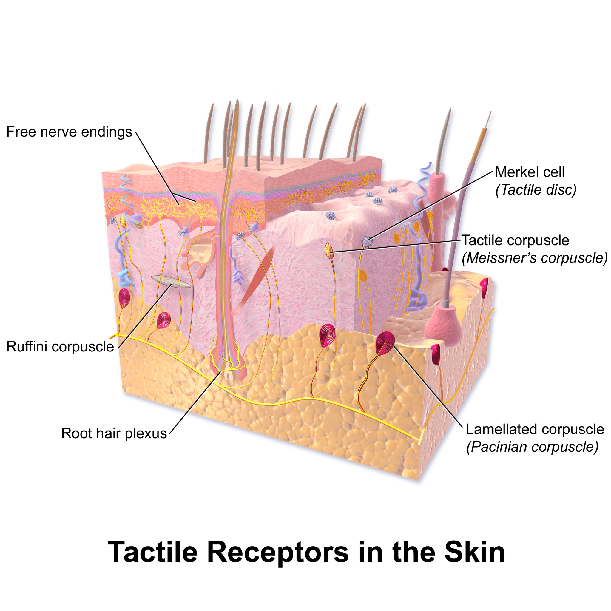
Touch is the ability to sense pressure, vibration, temperature, pain, and other tactile stimuli. These types of stimuli are detected by mechanoreceptors, thermoreceptors, and nociceptors all over the body, most noticeably in the skin. These receptors are especially concentrated on the tongue, lips, face, palms of the hands, and soles of the feet. Various types of tactile receptors in the skin are shown in Figure 8.7.2.
Vision
Vision (or sight) is the ability to sense light and see. The eye is the special sensory organ that collects and focuses light and forms images. The eye, however, is not sufficient for us to see. The brain also plays a necessary role in vision. Vision is our primary sense and more than 50 per cent of the cerebral cortex is devoted to processing visual information. A person with normal colour vision can differentiate between hundreds of thousands of different colours, hues, and shades.
How the Eye Works
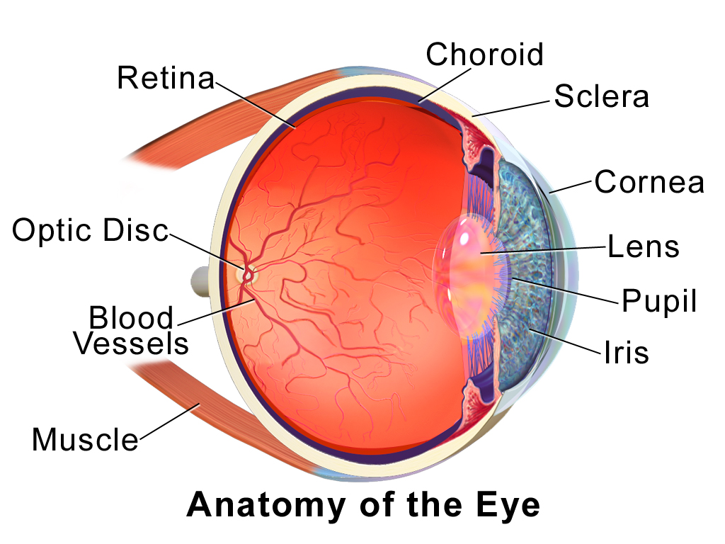
Figure 8.7.4 (below) shows the anatomy of the human eye in cross-section. The eye gathers and focuses light to form an image, and then changes the image to nerve impulses that travel to the brain. The eye’s functions are summarized in the following steps.
- Light passes first through the cornea, which is a clear outer layer that protects the eye and helps to focus the light by refracting (or bending) it.
- Next, light enters the interior of the eye through an opening called the pupil. The size of this opening is controlled by the coloured part of the eye (called the iris), which adjusts the size based on the brightness of the light. The iris causes the pupil to narrow in bright light and widen in dim light. Filling the space between the cornea and the iris is a semi-gelatinous fluid called aqueous humor and functions to maintain the shape of the eye.
- The light then passes through the lens, which refracts the light even more and focuses it on the retina at the back of the eye, as an inverted image. Sitting behind the lens is a gelatinous fluid called vitreous humor, which functions to maintain the shape of the eye.
- The retina contains two types of photoreceptors: rod and cone cells . Rod cells, which are found mainly in all areas of the retina other than the very center, are particularly sensitive to low levels of light. Cone cells, which are found mainly in the center of the retina, are sensitive to light of different colours, and allow colour vision. The rods and cones convert the light that strikes them to nerve impulses.
- The nerve impulses from the rods and cones travel to the optic nerve via the optic disc (also known as the optic nerve), which is a circular area at the back of the eye where the optic nerve connects to the retina.
Colour Vision
Humans have colour vision because we have three types of cone cells: blue, green and red. Each of these types of cone cell detects a specific wavelength of light, for which they are named. The combined stimulus is then perceived as a specific colour, based on the ratio of the amount stimulus coming from each of the three types of cone cells. Do you know what else uses these same three pieces of information to communicate colour? Your computer monitor! When working in a creative program, such as Paint, these three reference points of red (R), green (G), and blue (B), can be used to create any of the million colours the human eye can perceive, as illustrated in Figure 8.7.5. Take a look at each of the numerical values for red, green, and blue and what colour their combined values create:
Figure 8.7.5 RGB colours.
Role of the Brain in Vision
The optic nerves from both eyes meet and cross just below the bottom of the hypothalamus in the brain. The information from both eyes is sent to the visual cortex in the occipital lobe of the cerebrum, which is part of the cerebral cortex. The visual cortex is the largest system in the human brain, and is responsible for processing visual images. It interprets messages from both eyes and “tells” us what we are seeing.
Vision Problems

Vision problems are very common. Two of the most common are myopia and hyperopia, and they often start in childhood or adolescence. Another common problem, called presbyopia, occurs in most people, beginning in middle adulthood. In all three conditions, the eyes fail to focus images correctly on the retina, resulting in blurred vision.
Myopia
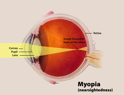
Myopia (or nearsightedness) occurs when the light that comes into the eye does not directly focus on the retina, but in front of it, as shown in Figure 8.7.7. As a result, distant objects may appear out of focus, but the focus of close objects is not affected. Myopia may occur because the eyeball is elongated from front to back, or because the cornea is too curved. Myopia can be corrected with the use of corrective lenses, either eyeglasses or contact lenses. Myopia can also be corrected by refractive surgery performed with a laser.
Hyperopia
Hyperopia (or farsightedness) happens when the light coming into the eye does not directly focus on the retina but behind it, as shown in Figure 8.7.8. This causes close objects to appear out of focus, but does not affect the focus of distant objects. Hyperopia may occur because the eyeball is too short from front to back, or because the lens is not curved enough. Hyperopia can be corrected through the use of corrective lenses or laser surgery.
Presbyopia
Presbyopia is a vision problem associated with aging, in which the eye gradually loses its ability to focus on close objects. The precise origin of presbyopia is not known for certain, but evidence suggests that the lens may become less elastic with age, causing the muscles that control the lens to lose power as people grow older. The first signs of presbyopia — eyestrain, difficulty seeing in dim light, problems focusing on small objects and fine print — are usually first noticed between the ages of 40 and 50. Most older people with this problem use corrective lenses to focus on close objects, because surgical procedures to correct presbyopia have not been as successful as those for myopia and hyperopia.
Hearing
Hearing is the ability to sense sound waves, and the ear is the organ that senses sound. Sound waves enter the ear through the ear canal and travel to the eardrum (see the diagram of the ear Figure 8.7.9). The sound waves strike the eardrum, and make it vibrate. The vibrations then travel through the three tiny bones (incus, malleus and stapes) of the middle ear, which amplify the vibrations. From the middle ear, the vibrations pass to the cochlea in the inner ear. The cochlea is a coiled tube filled with liquid. The liquid moves in response to the vibrations, causing tiny hair cells(which are mechanoreceptors) lining the cochlea to bend. In response, the hair cells send nerve impulses to the auditory nerve, which carries the impulses to the brain. The brain interprets the impulses and “tells” us what we are hearing.
Balance
The ears are also responsible for the sense of balance. Balance is the ability to sense and maintain an appropriate body position. The semicircular canals inside the ear (see the figure above) contain fluid that moves when the head changes position. Tiny hairs lining the semicircular canals sense movement of the fluid. In response, they send nerve impulses to the vestibular nerve, which carries the impulses to the brain. The brain interprets the impulses and sends messages to the peripheral nervous system, which triggers contractions of skeletal muscles as needed to maintain balance.
Taste and Smell
Taste and smell are both abilities to sense chemicals, so both taste and olfactory (odor) receptors are chemoreceptors. Both types of chemoreceptors send nerve impulses to the brain along sensory nerves, and the brain “tells” us what we are tasting or smelling.
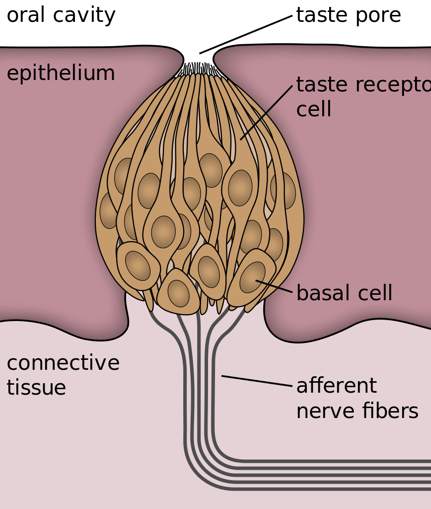
Taste receptors are found in tiny bumps on the tongue called taste buds.You can see a diagram of a taste receptor cell and related structures in Figure 8.7.10. Taste receptor cells make contact with chemicals in food through tiny openings called taste pores. When certain chemicals bind with taste receptor cells, it generates nerve impulses that travel through afferent nerves to the CNS. There are separate taste receptors for sweet, salty, sour, bitter, and meaty tastes. The meaty — or savory — taste is called umami.
Feature: Human Biology in the News
The most common cause of blindness in the Western hemisphere is age-related macular degeneration (AMD). Approximately 1.4 million people in Canada have this type of blindness, and 196 million people are affected worldwide and is expected to increase to 288 millions people by the year 2040. At present, there is no cure for AMD. The disease occurs with the death of a layer of cells called retinal pigment epithelium, which normally provides nutrients and other support to the macula of the eye. The macula is an oval-shaped pigmented area near the center of the retina that is specialized for high visual acuity and has the retina’s greatest concentration of cones. When the epithelial cells die and the macula is no longer supported or nourished, the macula also starts to die. Patients experience a black spot in the center of their vision, and as the disease progresses, the black spot grows outward. Patients eventually lose the ability to read and even to recognize familiar faces before developing total blindness.
In 2016, a landmark surgery was performed as a trial on a patient with severe AMD. In the first ever operation of its kind, Dr. Pete Coffey of the University of London implanted a tiny patch of cells behind the retina in each of the patient’s eyes. The cells were retinal pigmented epithelial cells that had been grown in a lab from stem cells, which are undifferentiated cells that can develop into other cell types. Within six months of the operation, the new cells were still surviving, and the doctor was hopeful that the patient’s vision loss would stop and even be reversed. At that point, several other operations had already been planned to test the new procedure. If these cases are a success, Dr. Coffey predicts that the surgery will become as routine as cataract surgery, and that it will prevent millions of patients from losing their vision.
8.7 Summary
- The human body has two major types of senses: special senses and general senses. Special senses have specialized sense organs and include vision (eyes), hearing (ears), balance (ears), taste (tongue), and smell (nasal passages). General senses are all associated with touch and lack special sense organs. Touch receptors are found throughout the body, but particularly in the skin.
- All senses depend on sensory receptor cells to detect sensory stimuli and transform them into nerve impulses. Types of sensory receptors include mechanoreceptors (mechanical forces), thermoreceptors (temperature), nociceptors (pain), photoreceptors (light), and chemoreceptors (chemicals).
- Touch is the ability to sense pressure, vibration, temperature, pain, and other tactile stimuli. The skin includes several different types of touch receptor cells.
- Vision is the ability to sense light and see. The eye is the special sensory organ that collects and focuses light, forms images, and changes them to nerve impulses. Optic nerves send information from the eyes to the brain, which processes the visual information and “tells” us what we are seeing.
- Common vision problems include myopia (nearsightedness), hyperopia (farsightedness), and presbyopia (age-related decline in close vision). Vision problems can be corrected with lenses (eyeglasses or contacts) or — in many cases — with laser surgery.
- Hearing is the ability to sense sound waves, and the ear is the organ that senses sound. It changes sound waves to vibrations that trigger nerve impulses, which travel to the brain through the auditory nerve. The brain processes the information and “tells” us what we are hearing.
- The ear is also the organ responsible for the sense of balance, which is the ability to sense and maintain an appropriate body position. The ears send impulses about head position to the brain, which sends messages to skeletal muscles via the peripheral nervous system. The muscles respond by contracting to maintain balance.
- Taste and smell are both abilities to sense chemicals. Taste receptors in taste buds on the tongue sense chemicals in food, while olfactory receptors in the nasal passages sense chemicals in the air. Sense of smell contributes significantly to sense of taste.
8.7 Review Questions
-
- Compare and contrast special senses and general senses.
- What are sensory receptors?
-
- Describe the range of tactile stimuli detected in the sense of touch.
- Explain how the eye collects and focuses light to form an image, and how it converts it to nerve impulses.
- Identify two common vision problems,along with their causes and their effects on vision.
-
- Explain how structures of the ear collect and amplify sound waves and transform them to nerve impulses.
- What role does the ear play in balance? Which structures of the ear are involved in balance?
- Describe two ways that the body senses chemicals. What are the special sense organs involved in these senses?
- Explain why your skin can detect different types of stimuli, such as pressure and temperature.
- Is sensory information sent to the central nervous system via efferent or afferent nerves?
- Identify a mechanoreceptor used in two different human senses. Describe the type of mechanical stimuli that each detects.
- If a person is blind, but their retina is functioning properly, where do you think the damage might be? Explain your answer.
- When you see colours, what receptor cells are activated? Where are these receptors located? What lobe of the brain is primarily used to process visual information?
- The auditory nerve carries _______________.
-
-
- smell information
- taste information
- balance information
- sound information
-
8.7 Explore More
What color is Tuesday? Exploring synesthesia – Richard E. Cytowic, TED-Ed, 2013.
What Is Vertigo & Why Do We Get It?, Seeker, 2016.
How do animals see in the dark? – Anna Stöckl, TED-Ed, 2016.
What are those floaty things in your eye? – Michael Mauser, TED-Ed, 2014.
Attributions
Figure 8.7.1
Bee Stereogram by Be Mosaic on Flickr is used under a CC BY-NC-SA 2.0 (https://creativecommons.org/licenses/by-nc-sa/2.0/) license.
Figure 8.7.2
Skin_TactileReceptors by BruceBlaus on Wikimedia Commons is used under a CC BY 3.0 (https://creativecommons.org/licenses/by/3.0) license.
Figure 8.7.3
Macro shot photograph of someone’s right eye [photo] by Jordan Whitfield on Unsplash is used under the Unsplash License (https://unsplash.com/license).
Figure 8.7.4
EyeAnatomy_01 by BruceBlaus on Wikimedia Commons is used under a CC BY 3.0 (https://creativecommons.org/licenses/by/3.0) license.
Figure 8.7.5
RGB colours [screenshots] from Microsoft Paint.
Figure 8.7.6
Through the reading glasses [photo] by Dmitry Ratushny on Unsplash is used under the Unsplash License (https://unsplash.com/license).
Figure 8.7.7
Myopia_Diagram by National Eye Institute/ National Institutes of Health on Wikimedia Commons is used under a CC BY 2.0 (https://creativecommons.org/licenses/by/2.0) license.
Figure 8.7.8
Hyperopia by National Institute of Health/NIH on Wikimedia Commons is in the public domain (https://en.wikipedia.org/wiki/Public_domain).
Figure 8.7.9
AnatomyHumanEar by unknown author from Occupational Safety & Health Administration on Wikimedia Commons is in the public domain (https://en.wikipedia.org/wiki/Public_domain).
Figure 8.7.10
Taste_bud_2_eng.svg by Jonas Töle on Wikimedia Commons is used under a CC0 1.0 Universal Public Domain Dedication license (https://creativecommons.org/publicdomain/zero/1.0/deed.en).
Figure 8.7.11
Head_olfactory_nerve by Patrick.lynch, medical illustrator on Wikimedia Commons is used under a CC BY 2.5 (https://creativecommons.org/licenses/by/2.5/deed.en) license.
References
Age-Related Macular Degeneration. (n.d.). WebMD. https://www.webmd.com/eye-health/macular-degeneration/age-related-macular-degeneration-overview#3 (Reviewed by Alan Kozarsky, MD on October 26, 2019)
Blausen.com Staff. (2014). Medical gallery of Blausen Medical 2014. WikiJournal of Medicine 1 (2). DOI:10.15347/wjm/2014.010. ISSN 2002-4436.
da Cruz, L., Fynes, K., Georgiadis, O. et al. (2018, March 19). Phase 1 clinical study of an embryonic stem cell–derived retinal pigment epithelium patch in age-related macular degeneration. Natural Biotechnology, 36, 328–337. https://doi.org/10.1038/nbt.4114
File:Eye Diagram without text.gif. (2018, February 9). Wikimedia Commons. https://commons.wikimedia.org/w/index.php?title=File:Eye_Diagram_without_text.gif&oldid=286008241 (original image from National Eye Institute – modified by User:Nordelch) [public domain (https://en.wikipedia.org/wiki/Public_domain)]
Occupational Health and Safety Administration. (n.d.). Figure 7. Anatomy of the human ear [diagram]. In OSHA Technical Manual (Section III, Chapter 5 – Noise). United States Department of Labour [online]. https://www.osha.gov/dts/osta/otm/new_noise/
Seeker. (2016, March 18). What is vertigo & why do we get it? YouTube. https://www.youtube.com/watch?v=UL8YSLhqa5U&feature=youtu.be
TED-Ed. (2013, June 10). What color is Tuesday? Exploring synesthesia – Richard E. Cytowic. YouTube. https://www.youtube.com/watch?v=rkRbebvoYqI&feature=youtu.be
TED-Ed. (2014, December 1). What are those floaty things in your eye? – Michael Mauser. YouTube. https://www.youtube.com/watch?v=Y6e_m9iq-4Q&feature=youtu.be
TED-Ed. (2016, August 25). How do animals see in the dark? – Anna Stöckl. YouTube. https://www.youtube.com/watch?v=t3CjTU7TaNA&feature=youtu.be
A diagram or computer-generated image giving a three-dimensional representation of a solid object or surface.
A sense, such as vision or hearing, that has special sense organs that gather sensory information and change it into nerve impulses.
A sense which lacks specialized sensory organs and is monitored instead by sensory receptors all over the body, such as the sense of touch.
Specialized nerve cell that responds to a particular type of stimulus such as light or chemicals by generating a nerve impulse.
A type of sensory receptor that responds to mechanical forces.
A type of sensory receptor that senses temperature.
A type of sensory receptor that responds to pain.
A type of sensory receptor that responds to light.
A type of sensory receptor that responds to presence of chemicals.
The ability to sense pressure, vibration, temperature, pain, and other tactile stimuli.
The transparent front part of the eye that covers the iris, pupil, and anterior chamber.
A hole located in the center of the iris of the eye that allows light to strike the retina.
A thin, annular structure in the eye, responsible for controlling the diameter and size of the pupil and thus the amount of light reaching the retina.
A transparent watery fluid similar to plasma, but containing low protein concentrations. It is secreted from the ciliary epithelium, a structure supporting the lens.
A transparent biconvex structure in the eye that, along with the cornea, helps to refract light to be focused on the retina.
The clear gel that fills the space between the lens and the retina of the eyeball of humans and other vertebrates. It is often referred to as the vitreous humor or simply "the vitreous".
A layer at the back of the eyeball containing cells that are sensitive to light and that trigger nerve impulses that pass via the optic nerve to the brain, where a visual image is formed.
Photoreceptor cells in the retina of the eye that can function in lower light than the other type of visual photoreceptor, cone cells. Rods are usually found concentrated at the outer edges of the retina and are used in peripheral vision.
One of the two types of photoreceptor cells that are in the retina of the eye which are responsible for color vision as well as eye color sensitivity; they function best in relatively bright light, as opposed to rod cells that work better in dim light.
The point of exit for ganglion cell axons leaving the eye. Because there are no rods or cones overlying the optic disc, it corresponds to a small blind spot in each eye. The ganglion cell axons form the optic nerve after they leave the eye.
A part of the brain that secretes hormones and connects the brain with the endocrine system.
A part of each hemisphere of the cerebrum that is dedicated almost solely to vision.
The largest part of the brain that controls conscious functions such as reasoning and sight.
The highly folded, thin outer layer of the cerebrum where most information processing in the brain takes place.
A vision problem in which distant objects are out of focus but close vision is unaffected; also called nearsightedness.
A vision problem in which close objects are out of focus but distant vision is unaffected; also called farsightedness.
The ability to sense sound waves.
A special sensory organ that collects and amplifies sound waves and information on body position and transforms them into nerve impulses that travel to the brain.
A coiled, fluid-filled tube in the inner ear that changes mechanical sound vibrations and positional information to nerve impulses that travel to the brain.
The ability to sense and maintain an appropriate body position.
Three tiny, fluid-filled tubes in your inner ear that help you keep your balance. When your head moves around, the liquid inside the semicircular canals sloshes around and moves the tiny hairs that line each canal.
The perception produced or stimulated when a substance in the mouth reacts chemically with taste receptor cells located on taste buds in the oral cavity, mostly on the tongue.
A chemoreception (ability to detect the presence of certain chemicals) that, through the sensory olfactory system, forms the perception of smell.
A small structure on the tongue containing chemoreceptor cells that sense chemicals in food.
Small openings in the tongue epithelium, which allow parts of the food dissolved in saliva come into contact with taste receptors.
An undifferentiated cell that can develop into specialized types of cells.
The ability to sense light and see; also called sight.
Farsightedness caused by loss of elasticity of the lens of the eye, occurring typically in middle and old age.
