14.2 Introduction to the Cardiovascular System
Created by CK-12 Foundation/Adapted by Christine Miller
Ant Hill or Plumbing System?
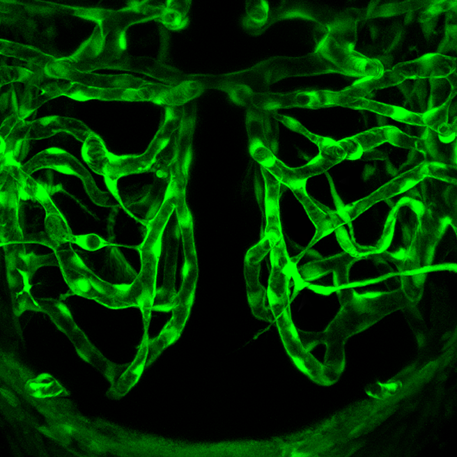
What do you think the picture in Figure 14.2.1 shows? Is it a maze of underground passageways in an ant hill? A network of interconnected pipes in a complex plumbing system? The picture actually shows something that, like ant tunnels and plumbing pipes, functions as a transportation system. It shows a network of blood vessels, which are part of the cardiovascular system.
What is the Cardiovascular System?
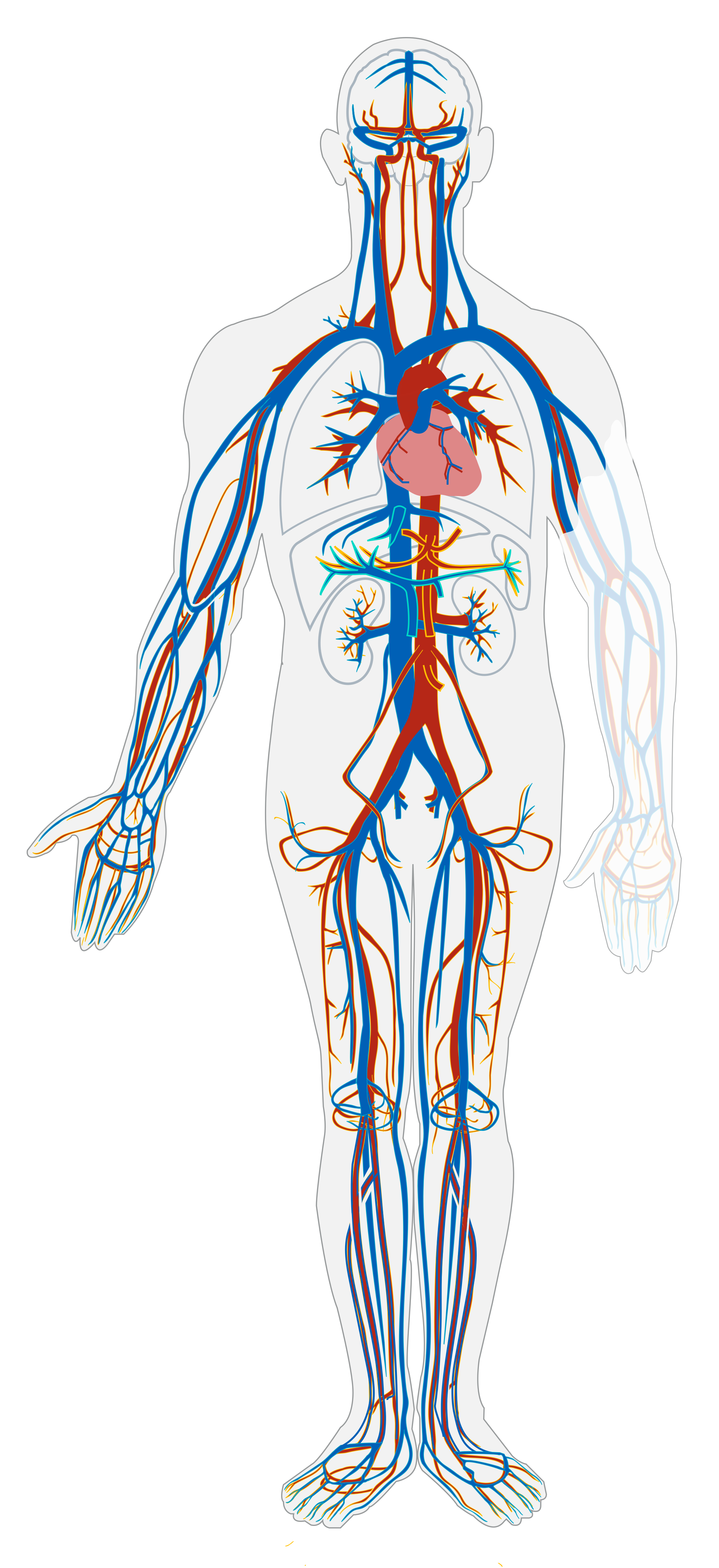
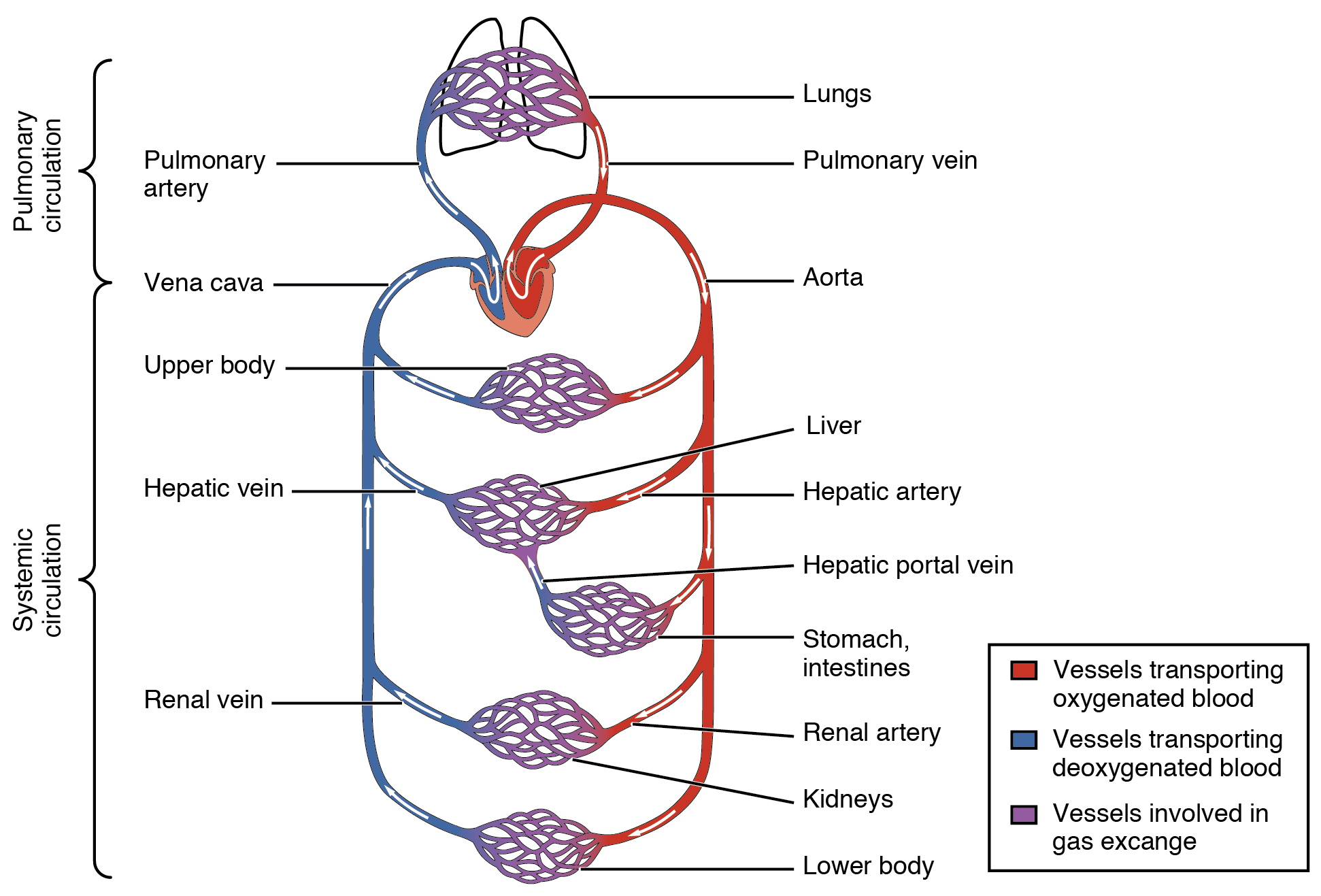
The cardiovascular system, also called the circulatory system, is the organ system that transports materials to and from all the cells of the body. The materials carried by the cardiovascular system include oxygen from the lungs, nutrients from the digestive system, hormones from glands of the endocrine system, and waste materials from cells throughout the body. Transport of these and many other materials is necessary to maintain homeostasis of the body. The main components of the cardiovascular system are the heart, blood vessels, and blood. Each of these components is shown in Figure 14.2.2 and introduced below.
Heart
The heart is a muscular organ in the chest. It consists mainly of cardiac muscle tissue, and it pumps blood through blood vessels by repeated, rhythmic contractions. As shown in Figure 14.2.3, the heart has four inner chambers: a right atrium and ventricle, and a left atrium and ventricle. On each side of the heart, blood is pumped from the atrium to the ventricle below it, and from the ventricle out of the heart. The heart also contains several valves that allow blood to flow only in the proper direction through the heart.
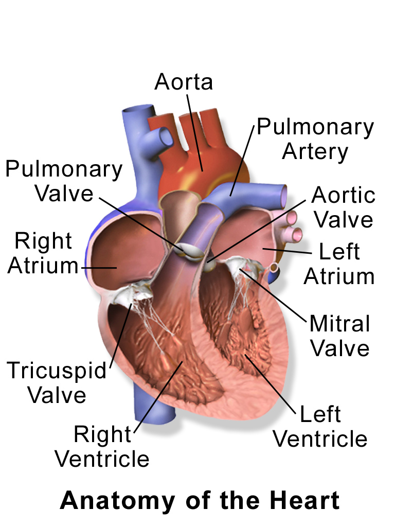
As you may have noticed, the Figure 14.2.3 diagram labels the right side of the heart on the left side of the diagram, and vice versa. This is because it is assumed that in this diagram, the heart appears as if the patient was facing us – the patient’s left side is on our right side!
Unlike skeletal muscle, cardiac muscle routinely contracts without stimulation by the nervous system. Specialized cardiac muscle cells send out electrical impulses that stimulate the contractions. As a result, the atria and ventricles normally contract with just the right timing to keep blood pumping efficiently through the heart.
Blood Vessels
The blood vessels of the cardiovascular system are like a network of interconnected, one-way roads that range from superhighways to back alleys. Like a network of roads, the blood vessels are tasked with allowing the transport of materials from one place to another. There are three major types of blood vessels: arteries, veins, and capillaries. They are illustrated in Figure 14.2.4 and described below.
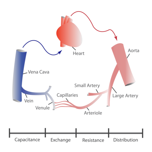
- Arteries are blood vessels that carry blood away from the heart (except for the arteries that actually supply blood to the heart muscle). Most arteries carry oxygen-rich blood, and one of their main functions is distributing oxygen to tissues throughout the body. The smallest arteries are called arterioles.
- Veins are blood vessels that carry blood toward the heart. Most veins carry deoxygenated blood. The smallest veins are called venules.
- Capillaries are the smallest blood vessels, and they connect arterioles and venules. As they pass through tissues, they exchange substances (including oxygen) with cells.
Two Circulations
Cells throughout the body need a constant supply of oxygen. They get oxygen from capillaries in the systemic circulation. The systemic circulation is just one of two interconnected circulations that make up the human cardiovascular system. The other circulation is the pulmonary system, which is where blood picks up oxygen to carry to cells. It takes blood about 20 seconds to make one complete transit through both circulations (see Figure 14.2.5).
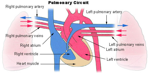
Pulmonary Circuit
The pulmonary circuit involves only the heart, the lungs, and the major blood vessels that connect them (illustrated in Figure 14.2.6). Blood moves through the pulmonary circuit from the heart, to the lungs, and then back to the heart again, becoming oxygenated in the process. Specifically, the right ventricle of the heart pumps deoxygenated blood into the right and left pulmonary arteries. These arteries carry the blood to the right and left lungs, respectively. Oxygenated blood then returns from the right and left lungs through the two right and two left pulmonary veins. All four pulmonary veins enter the left atrium of the heart.
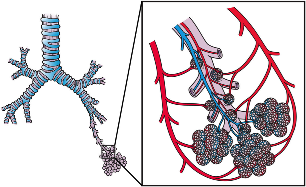
What happens to the blood while it is in the lungs? It passes through increasingly smaller arteries, and finally through capillary networks surrounding the alveoli (see Figure 14.2.7). This is where gas exchange takes place. The deoxygenated blood in the capillaries picks up oxygen from the alveoli, and gives up carbon dioxide to the alveoli. As a result, the blood returning to the heart in the pulmonary veins is almost completely saturated with oxygen.
Systemic Circulation
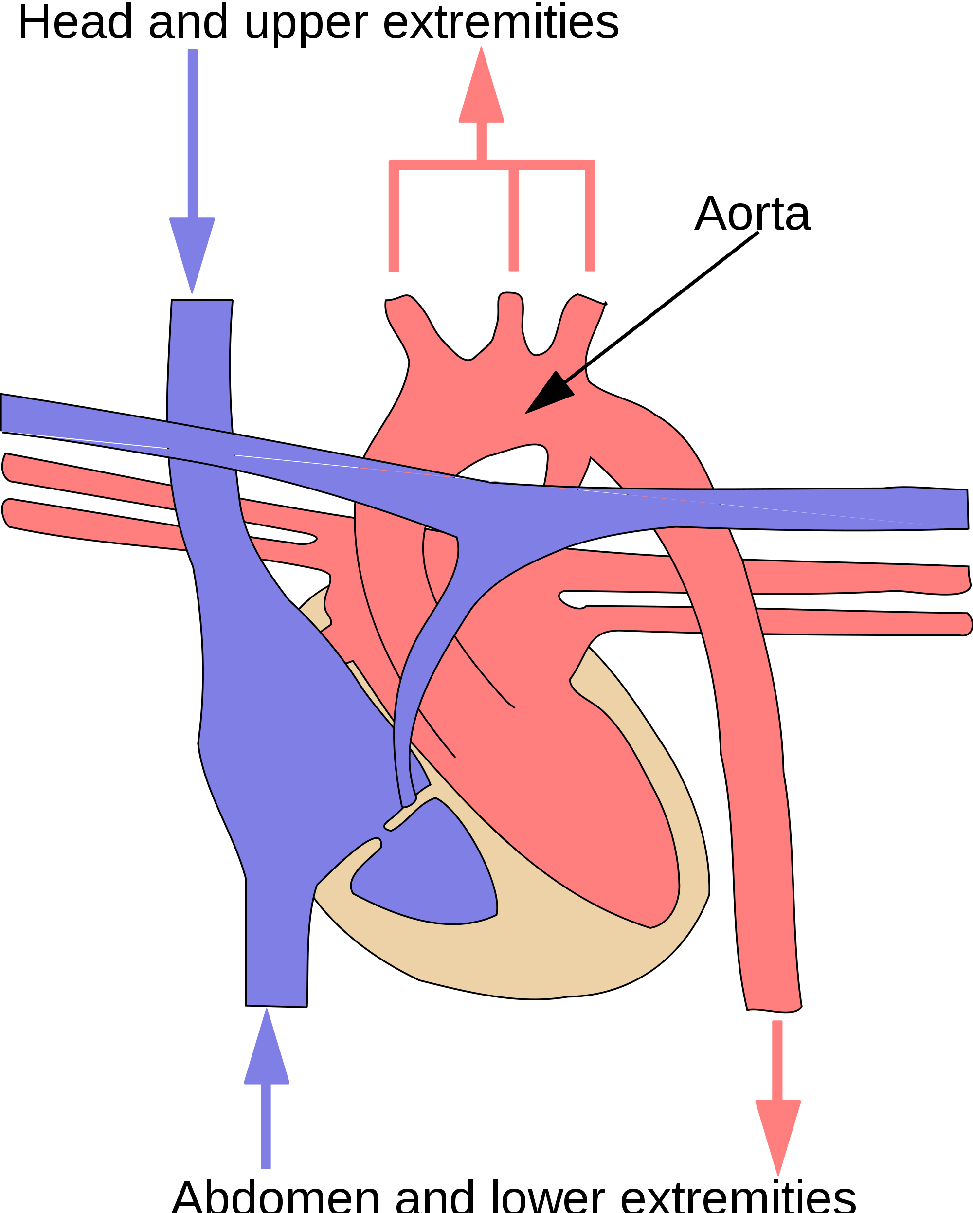
The oxygenated blood that enters the left atrium of the heart in the pulmonary circulation then passes into the systemic circuit. This is the part of the cardiovascular system that transports blood to and from all of the tissues of the body to provide oxygen and nutrients, and to pick up wastes. It consists of the heart and blood vessels that supply the metabolic needs of all the cells in the body, including those of the heart and lungs.
As shown in Figure 14.2.8, in the systemic circulation, the left atrium pumps oxygenated blood to the left ventricle, which pumps the blood directly into the aorta, the body’s largest artery. Major arteries branching off the aorta carry the blood to the head and upper extremities. The aorta continues down through the abdomen and carries blood to the abdomen and lower extremities. The blood then returns to the heart through the network of increasingly larger veins of the systemic circulation. All of the returning blood eventually collects in the superior vena cava (upper body) and inferior vena cava (lower body), which empty directly into the right atrium of the heart.
Blood
Blood is a fluid connective tissue that circulates throughout the body in blood vessels by the pumping action of the heart. Blood carries oxygen and nutrients to all the body’s cells, and it carries carbon dioxide and other wastes away from the cells to be excreted. Blood also transports many other substances, defends the body against infection, repairs body tissues, and controls the body’s pH, among other functions.
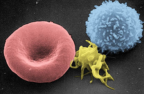
The fluid part of blood is called plasma. It is a yellowish, watery liquid that contains many dissolved substances and blood cells. Types of blood cells in plasma include red blood cells, white blood cells, and platelets, all of which are illustrated in the photomicrograph (Figure 14.2.9) and described below.
- Erythrocytes (red blood cells) have the main function of carrying oxygen in the blood. Red blood cells consist mostly of hemoglobin, a protein containing iron that binds with oxygen.
- Leukocytes (white blood cells) are far fewer in number than red blood cells. They defend the body in various ways. White blood cells called phagocytes, for example, swallow and destroy pathogens, dead cells, and other debris in the blood.
- Thrombocytes (platelets) are cell fragments involved in blood clotting. They stick to tears in blood vessels and to each other, forming a plug at the site of injury. They also release chemicals that are needed for clotting to occur.
14.2 Summary
- The cardiovascular system is the organ system that transports materials to and from all the cells of the body. The main components of the cardiovascular system are the heart, blood vessels, and blood.
- The heart is a muscular organ in the chest that consists mainly of cardiac muscle and pumps blood through blood vessels by repeated, rhythmic contractions. The heart has four chambers through which blood flows, and valves that keep blood flowing in just one direction.
- Blood vessels carry blood throughout the body. Major types of blood vessels are arteries (which mainly carry blood away from the heart), veins (which carry blood toward the heart), and capillaries (which exchange substances between the blood and cells of the body).
- The cardiovascular system has two interconnected circulations. The pulmonary circuit carries blood between the heart and lungs, where blood is oxygenated. The systemic circuit carries blood between the heart and the rest of the body, where it delivers oxygen.
- Blood is a fluid connective tissue that circulates throughout the body in blood vessels. It consists of a liquid part — called plasma — which contains many dissolved substances, and cells, including erythrocytes, leukocytes and thrombocytes.
14.2 Review Questions
- Describe the heart and how it functions.
- Compare and contrast the pulmonary and systemic circulations.
-
- What is blood? What are its chief constituents?
- Name three different types of substances transported by the cardiovascular system.
- Explain why the heart and lungs need blood from the systemic circulation.
- Do blood vessels carrying deoxygenated blood from the body back to the heart get increasingly larger or smaller?
14.2 Explore More
How the heart actually pumps blood – Edmond Hui, TED-Ed, 2014.
Circulatory & Respiratory Systems – CrashCourse Biology #27, CrashCourse, 2012.
The Heart and Circulatory System – How They Work, Mayo Clinic, 2013.
Attributions
Figure 14.2.1
Brain vascular formation [photo] by Liulin Du/ Chen (The National Cancer Institute at Frederick) on PLOS Biology is used under a CC BY 4.0 license.
Figure 14.2.2
Circulatory_System_no_tags.svg by Mariana Ruiz Villarreal [LadyofHats] on Wikimedia Commons is released into the public domain (https://en.wikipedia.org/wiki/Public_domain).
Figure 14.2.3
Blausen_0462_HeartAnatomy by BruceBlaus on Wikimedia Commons is used under a CC BY 3.0 (https://creativecommons.org/licenses/by/3.0)
Figure 14.2.4
Structure and functions of the different types of blood vessels by CK-12 Foundation is used under a CC BY-NC 3.0 (https://creativecommons.org/licenses/by-nc/3.0/) license.
Figure 14.2.5
2101_Blood_Flow_Through_the_Heart by OpenStax College on Wikimedia Commons is used under a CC BY 3.0 (https://creativecommons.org/licenses/by/3.0) license.
Figure 14.2.6
Illu_pulmonary_circuit by Arcadian from National Cancer Institute/ SEER Training on Wikimedia Commons is in the the public domain (https://en.wikipedia.org/wiki/Public_domain).
Figure 14.2.7
Pulmonary_Blood_Circulation by Artwork by Holly Fischer from Open Michigan (Respiratory Tact Slide 20) on Wikimedia Commons is used under a CC BY 3.0 (https://creativecommons.org/licenses/by/3.0) license.
Figure 14.2.8
systemic_circuit.svg by Surachit on Wikimedia Commons is used under a CC BY-SA 3.0 (http://creativecommons.org/licenses/by-sa/3.0/) license. (Derivative work based on SEER Training by NCI/ U.S. Government).
Figure 14.2.9
Red_White_Blood_cells by Electron Microscopy Facility at The National Cancer Institute at Frederick (NCI-Frederick) on Wikimedia Commons is in the public domain (https://en.wikipedia.org/wiki/Public_domain).
References
Blausen.com Staff. (2014). Medical gallery of Blausen Medical 2014. WikiJournal of Medicine 1 (2). DOI:10.15347/wjm/2014.010. ISSN 2002-4436
Betts, J. G., Young, K.A., Wise, J.A., Johnson, E., Poe, B., Kruse, D.H., Korol, O., Johnson, J.E., Womble, M., DeSaix, P. (2013, June 19). Figure 20.2 Cardiovascular circulation [digital image]. In Anatomy and Physiology (Section 7.3). OpenStax. https://openstax.org/books/anatomy-and-physiology/pages/20-1-structure-and-function-of-blood-vessels
Brainard, J/ CK-12 Foundation. (2016). Figure 4 Diagram represents the structure and functions of the different types of blood vessels in the cardiovascular system [digital image]. In CK-12 College Human Biology (Section 16.2) [online Flexbook]. CK12.org. https://www.ck12.org/book/ck-12-college-human-biology/section/16.2/
CrashCourse. (2012, July 30). Circulatory & respiratory systems – CrashCourse Biology #27. YouTube. https://www.youtube.com/watch?v=9fxm85Fy4sQ&feature=youtu.be
Du, J. (2012, August). Brain vasculature formation [digital image]. PLoS Biology, 10(8): ev10.i08. https://doi.org/10.1371/image.pbio.v10.i08 © Chen.
Mayo Clinic. (2013). The heart and circulatory system – How they work. YouTube. https://www.youtube.com/watch?v=CWFyxn0qDEU&t=1s
TED-Ed. (2014, May 20). How the heart actually pumps blood – Edmond Hui. YouTube. https://www.youtube.com/watch?v=ruM4Xxhx32U&feature=youtu.be
Refers to the body system consisting of the heart, blood vessels and the blood. Blood contains oxygen and other nutrients which your body needs to survive. The body takes these essential nutrients from the blood.
Two paired organs of the respiratory system in which gas exchange takes place between the blood and the atmosphere.
A body system including a series of hollow organs joined in a long, twisting tube from the mouth to the anus. The hollow organs that make up the GI tract are the mouth, esophagus, stomach, small intestine, large intestine, and anus. The liver, pancreas, and gallbladder are the solid organs of the digestive system.
A hormone is a signaling molecule produced by glands in multicellular organisms that target distant organs to regulate physiology and behavior.
The body system which acts as a chemical messenger system comprising feedback loops of the hormones released by internal glands of an organism directly into the circulatory system, regulating distant target organs. In humans, the major endocrine glands are the thyroid gland and the adrenal glands.
The ability of an organism to maintain constant internal conditions despite external changes.
A muscular organ in the chest that pumps blood through blood vessels when it contracts.
Involuntary, striated muscle found only in the walls of the heart; also called myocardium.
A hollow, tube-like structure through which blood flows in the cardiovascular system; vein, artery, or capillary.
A type of blood vessel that carries blood away from the heart and toward the lungs or body.
A type of blood vessel that carries blood toward the heart from the lungs or body.
The smallest type of blood vessel that connects arterioles and venules and that transfers substances between blood and tissues.
The part of the cardiovascular system that carries blood between the heart and lungs.
The part of the cardiovascular system that carries blood between the heart and body.
A body fluid in humans and other animals that delivers necessary substances such as nutrients and oxygen to the cells and transports metabolic waste products away from those same cells. In vertebrates, it is composed of blood cells suspended in blood plasma.
A measure of the acidity or basicity of aqueous or other liquid solutions. The term translates the values of the concentration of the hydrogen ion in a scale ranging from 0 and 14. In pure water, which is neutral (neither acidic nor alkaline), the concentration of the hydrogen ion corresponds to a pH of 7. A solution with a pH less than 7 is considered acidic; a solution with a pH greater than 7 is considered basic, or alkaline.
A straw-yellow fluid part of blood that contains many dissolved substances and blood cells.
A red blood cell that (in humans) is typically a biconcave disc without a nucleus. Erythrocytes contain the pigment hemoglobin, which imparts the red color to blood, and transport oxygen and carbon dioxide to and from the tissues.
An oxygen-binding protein containing iron that is the principal component of red blood cells.
a colorless cell that circulates in the blood and body fluids and is involved in counteracting foreign substances and disease; a white (blood) cell. There are several types, all amoeboid cells with a nucleus, including lymphocytes, granulocytes, monocytes, and macrophages.
Another term for platelet; a small colorless disk-shaped cell fragment without a nucleus, found in large numbers in blood and involved in clotting.
A form of connective tissue in which the matrix is in a liquid state. Examples include blood and lymph.
