8.5 Central Nervous System
Created by CK-12 Foundation/Adapted by Christine Miller
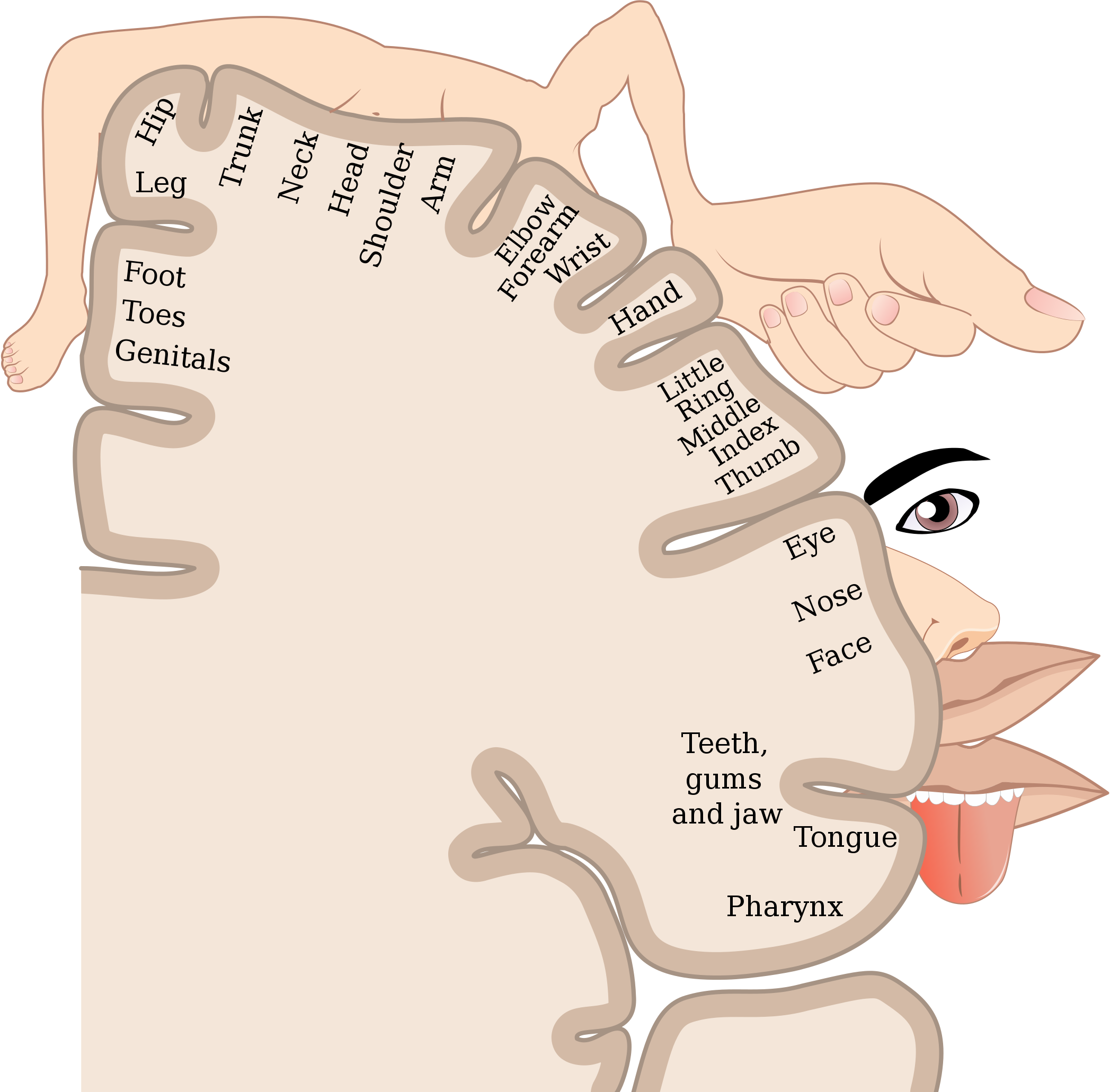
Homunculus
The very odd-looking drawing in Figure 8.5.1 is called a homunculus. The beige mass represents a cross-sectional wedge of the human brain, and the drawing shows some areas of the brain associated with different parts of the body. As you can see, larger areas of the brain in this region are associated with the hands, face, and tongue, as compared to the areas associated with the legs and feet. Given the importance of speech, manual dexterity, and face-to-face social interactions in human beings, it is not surprising that relatively large areas of the brain are needed to control these body parts. The brain is the most complex organ in the human body and part of the central nervous system.
What Is the Central Nervous System?
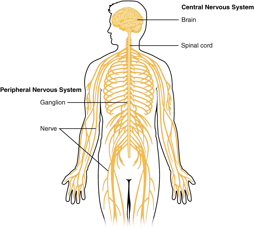
The central nervous system (CNS) is the part of the nervous system that includes the brain and spinal cord. The drawing below (Figure 8.5.2) shows the central nervous system as one of two main divisions of the total nervous system. The other main division is the peripheral nervous system (PNS). The CNS and PNS work together to control virtually all body functions.
The delicate nervous tissues of the central nervous system are protected by major physical and chemical barriers. Physically, the brain and spinal cord are surrounded by tough meninges, a three-layer protective sheath that also contains cushioning cerebrospinal fluid. The bones of the skull and spinal vertebrae also contribute to physically protecting the brain and spinal cord. Chemically, the brain and spinal cord are isolated from the circulation — and most toxins or pathogens in the blood — by the blood-brain barrier. The blood-brain barrier is a highly selective membrane formed of endothelial cells that separates the circulating blood from extracellular fluid in the CNS. The barrier allows water, certain gases, glucose, and some other molecules needed by the brain and spinal cord to cross from the blood into the CNS, while keeping out potentially harmful substances. These physical and chemical barriers make the CNS less susceptible to injury than the PNS. However, damage to the CNS is likely to have more serious consequences.
The Brain
The brain is the control center of not only the rest of the nervous system, but of the entire organism. The adult brain makes up only about 2% of the body’s weight, but it uses about 20% of the body’s total energy. The brain contains an estimated 100 billion neurons, and each neuron has thousands of synaptic connections to other neurons. The brain also has about the same number of neuroglia as neurons. No wonder the brain uses so much energy! In addition, the brain uses mostly glucose for energy. As a result, if the brain is deprived of glucose, it can lead to unconsciousness. The brain is able to store some glucose in the form of glycogen, but in much smaller amounts than are found in the liver and skeletal muscles.
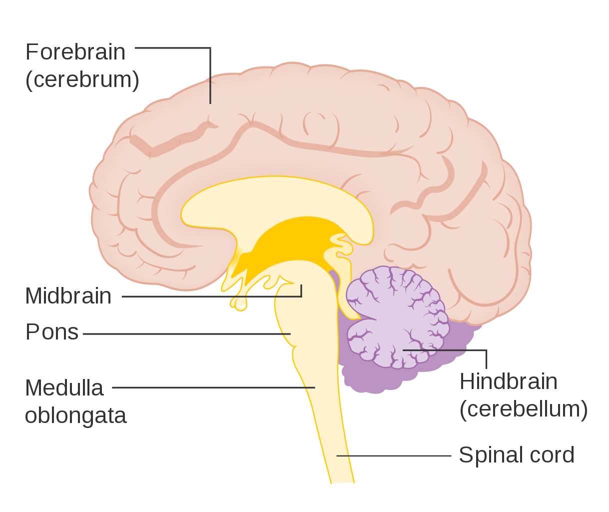
The brain controls such mental processes as reasoning, imagination, memory, and language. It also interprets information from the senses and commands the body to respond appropriately. It controls basic physical processes (such as breathing and heartbeat), as well as voluntary activities (such as walking and writing). The brain has three major regions: the hindbrain, the midbrain and the forebrain. These parts are shown in Figure 8.5.3 and described below.
The Hindbrain
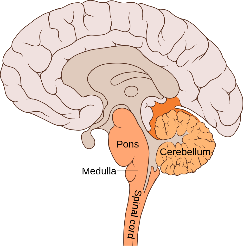
The hindbrain, which includes the cerebellum, medulla oblongata, and the pons. The hindbrain is the lowest part of the brain. It resembles a stalk and platform on which the cerebrum is perched. The components of the hindbrain connect the rest of the brain with the spinal cord and passes nerve impulses between the brain and spinal cord.
Cerebellum
The cerebellum is located just below the cerebrum and at the back of the brain behind the brain stem. It coordinates your voluntary movements, balance, and posture. Information from your inner ear, joints and muscles, and eyes are all knitted together in the cerebellum so that you have awareness of where you are in 3-dimensional space. Patients who have suffered damage to their cerebellum may suffer from balance disorder. In addition the cerebellum plays a major role in motor learning (like how to ride a bike or how to do a backflip on a trampoline) through trial and error. While traditionally, the cerebellum was thought to only be involved in motor functions, we now know that it also plays an important role in memory and learning.
Medulla Oblongata
The medulla oblongata makes up part of the brainstem and sits in front of and just below the cerebellum, at the very top of the spinal cord. It is responsible for control of heart rate, respiration rate and blood pressure, as well as reflexes such as vomiting, coughing, sneezing and swallowing.
Pons
The pons is located in front of the cerebellum and above the medulla oblongata. It has several functions, including receiving sensory information from the face, regulating rate and depth of breaths, as well as sleep cycles.
Midbrain
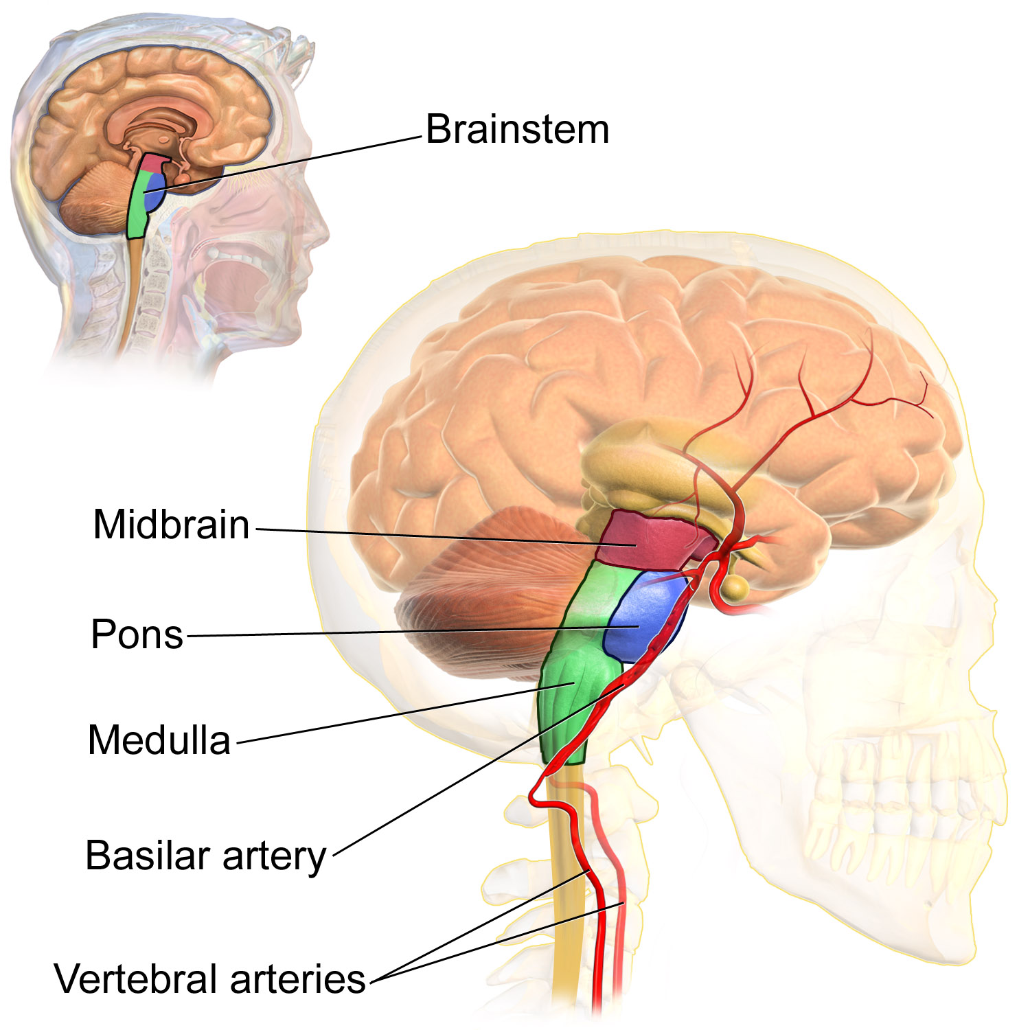
The midbrain is the topmost part of the brainstem and is the essential connection between and brain and spinal cord. The three main parts of the midbrain are the colliculi, the tegmentum, and the cerebral peduncles. These structures are all considered part of the brainstem, which consists of the medulla oblongata, the pons and the midbrain.
Reticular Activating System
The reticular activating system (RAS) is responsible for the sleep-wake cycle and wakefulness. It also regulates attention, ability to focus and arousal.
Forebrain
The forebrain is the anterior (forwardmost) part of the brain and includes the cerebrum, the thalamus and hypothalamus, hippocampus, amygdala, and limbic system. This portion of the brain is responsible for processing incoming sensory information, performing complex cognitive activities (speech, abstract thought, etc) and governing voluntary motor movements. The forebrain also controls body temperature, reproductive functions, eating, sleeping and the display of emotions.
Cerebrum
The cerebrum is the largest part of the brain. It controls conscious, intellectual functions. Among other things, it controls reasoning, language, memory, sight, touch, and hearing. When you read a book, play a video game, or recognize a classmate, you are using your cerebrum.
Hemispheres and Lateralization of the Cerebrum
The cerebrum is divided from front to back into two halves called the left and right hemispheres. The two hemispheres are connected by a thick bundle of axons, known as the corpus callosum, which lies deep within the brain. The corpus callosum is the main avenue of communication between the two hemispheres. It connects each point in the cerebrum to the mirror-image point in the opposite hemisphere.
The right and left hemispheres of the cerebrum are similar in shape, and most areas of cerebrum are found in both hemispheres. Some areas, however, show lateralization, or a concentration in one hemisphere or the other. In most people, for example, language functions are more concentrated in the left hemisphere, whereas abstract reasoning and visual-spatial abilities are more concentrated in the right hemisphere.
For reasons that are not yet clear, each hemisphere of the brain interacts primarily with the opposite side of the body. The left side of the brain receives messages from and sends commands to the right side of the body, and the right side of the brain receives messages from and sends commands to the left side of the body. Sensory nerves from the spinal cord to the brain and motor nerves from the brain to the spinal cord both cross the midline of the body at the level of the brain stem.
Lobes of the Cerebrum
Each hemisphere of the cerebrum is further divided into the four lobes shown in Figure 8.5.6 and described below.
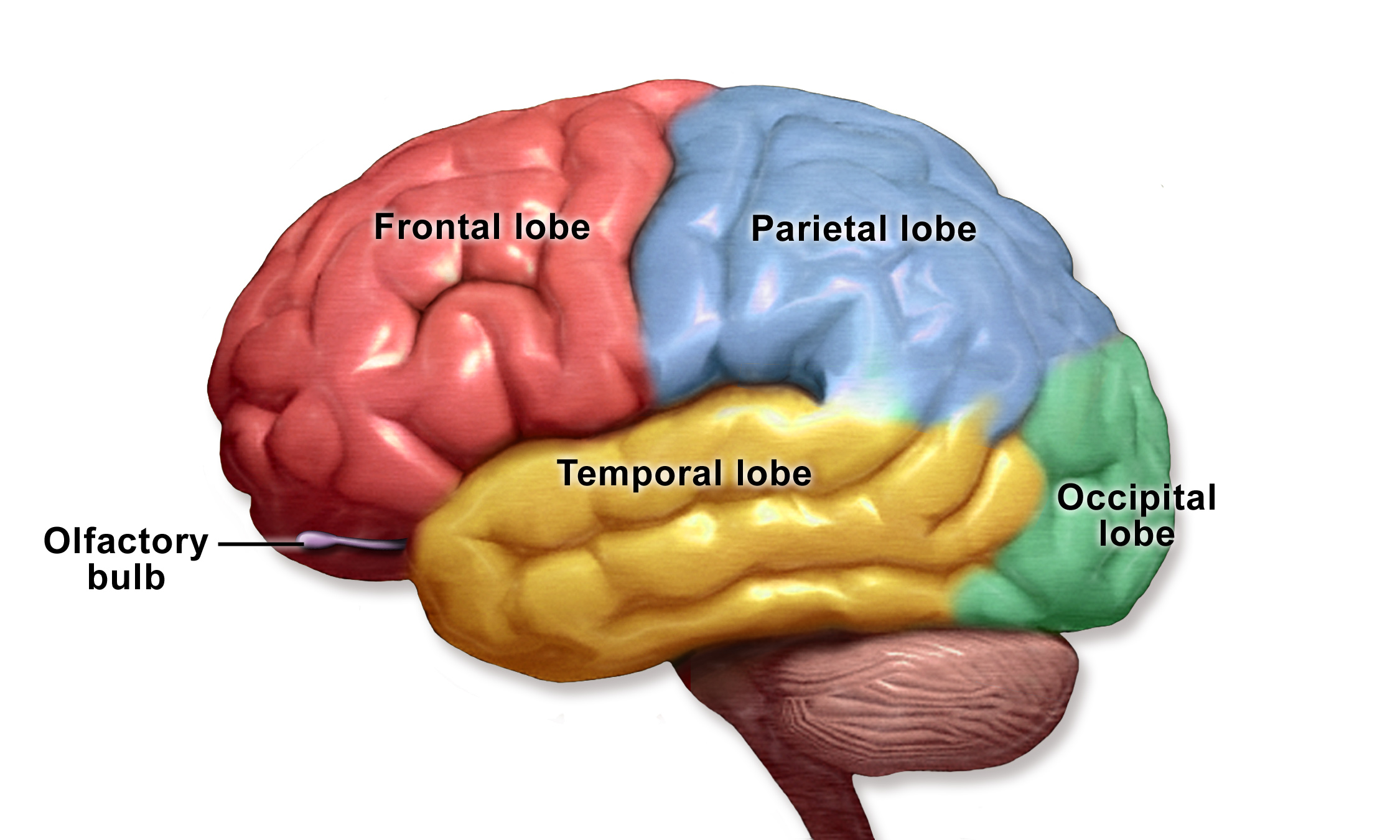
Each hemisphere of the cerebrum consists of four parts, called lobes. Each lobe is associated with particular brain functions. Just one function of each lobe is listed here.
- The frontal lobes are located at the front of the brain behind the forehead. The frontal lobes are associated with executive functions, such as attention, self-control, planning, problem solving, reasoning, abstract thought, language, and personality.
- The parietal lobes are located behind the frontal lobes at the top of the head. The parietal lobes are involved in sensation — including temperature, touch, and taste. Reading and arithmetic are also functions of the parietal lobes.
- The temporal lobes are located at the sides of the head below the frontal and parietal lobes. The temporal lobes enable hearing, the formation and retrieval of memories, and the integration of memories and sensations.
- The occipital lobes are located at the back of the head below the parietal lobes. The occipital lobes are the smallest of the four pairs of lobes. They are dedicated almost solely to vision.
Cerebral Cortex
Most of the information processing in the brain actually takes place in the cerebral cortex, a rind of gray matter and other tissues just a few millimetres thick that makes up the outer surface of the cerebrum in both hemispheres of the brain. The cerebral cortex has many folds in it, greatly increasing the amount of surface area of the brain that can fit within the skull. Because of all the folds in the human cerebral cortex, it has a surface area of about 2,500 cm2 (2.5 ft2). The size and importance of the cerebral cortex is far greater in the human brain than the brains of any other vertebrates, including nonhuman primates.
The Limbic System
The limbic system consists of the hypothalamus, thalamus, the hippocampus and amygdala. The structures and interacting areas of the limbic system are involved in motivation, emotion, learning, and memory.
Thalamus and Hypothalamus
Several structures are located deep within the brain and are important for communication between the brain and spinal cord (or the rest of the body). These structures include the hypothalamus and thalamus. The diagram below (Figure 8.5.7) shows where these structures are located in the brain. Like the two halves of the cerebrum, the hypothalamus and thalamus exist in two halves, one in each hemisphere.
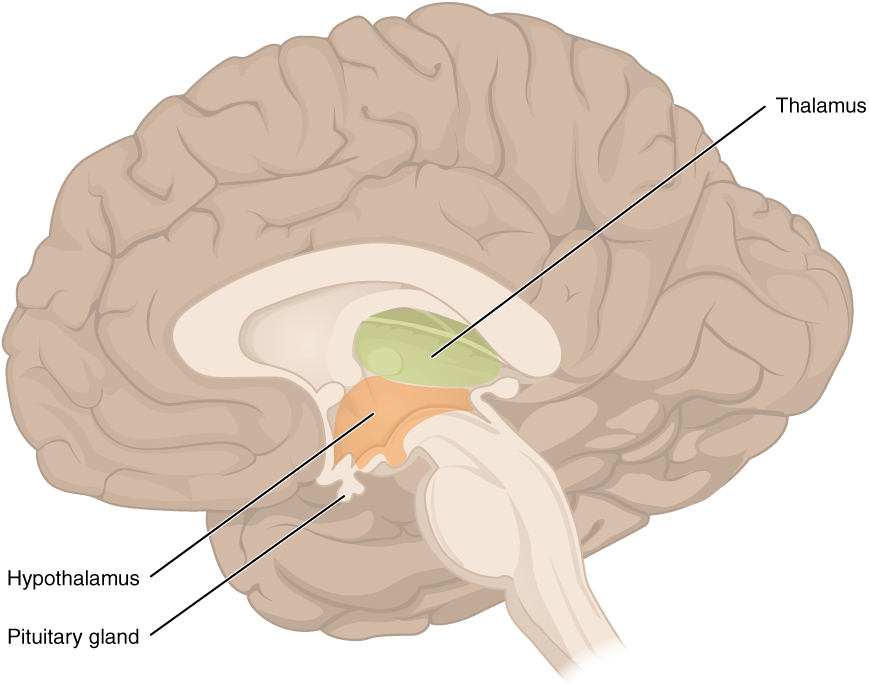
The hypothalamus is located just above the brain stem, and is about the size of an almond. The hypothalamus is responsible for certain metabolic processes and other activities of the autonomic nervous system, including body temperature, heart rate, hunger, thirst, fatigue, sleep, wakefulness, and circadian (24-hour) rhythms. The hypothalamus is also an important emotional center of the brain. The hypothalamus can regulate so many body functions because it responds to many different internal and external signals, including messages from the brain, light, steroid hormones, stress, and invading pathogens, among others.
One way the hypothalamus influences body functions is by synthesizing hormones that directly influence body processes. It synthesizes the hormone oxytocin, which stimulates uterine contractions during childbirth and the letdown of milk during lactation. It also synthesizes antidiuretic hormone, which stimulates the kidneys to reabsorb more water and excrete more concentrated urine. These two hormones are sent from the hypothalamus via a stalk-like structure called the infundibulum (see Figure 8.5.7) directly to the posterior (back) portion of the pituitary gland, which secretes them into the blood.
The main way the hypothalamus influences body functions is by controlling the pituitary gland, known as the master gland of the endocrine system. The hypothalamus synthesizes neurohormones called releasing factors that travel through the infundibulum directly to the anterior (front) part of the pituitary gland. The releasing factors generally either stimulate or inhibit the secretion of anterior pituitary hormones, most of which control other glands of the endocrine system.
The thalamus, which is located near the hypothalamus (see Figure 8.5.7), is a major hub for information traveling back and forth between the spinal cord and cerebrum. It relays sensory signals to the cerebral cortex and motor signals to the spinal cord. It is also involved in the regulation of consciousness, sleep, and alertness.
Hippocampus and Amygdala
The hippocampus is a complex structure embedded deep in the temporal lobe. It plays a major role in learning and memory and contributes to regulation of motivation and emotion. The amygdala is the part of the brain responsible for formation and storage of memories associated with emotional events.
Watch “The Limbic System” by Soton Brain Hub to learn about the location and functions of the limbic system.
The Limbic System, Soton Brain Hub, 2016.
Spinal Cord
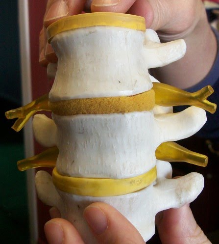
The spinal cord is a long, thin, tubular bundle of nervous tissues that extends from the brain stem and continues down the center of the back to the pelvis. It is highlighted in yellow in Figure 8.5.8. The spinal cord is enclosed within, but is shorter than, the vertebral column.
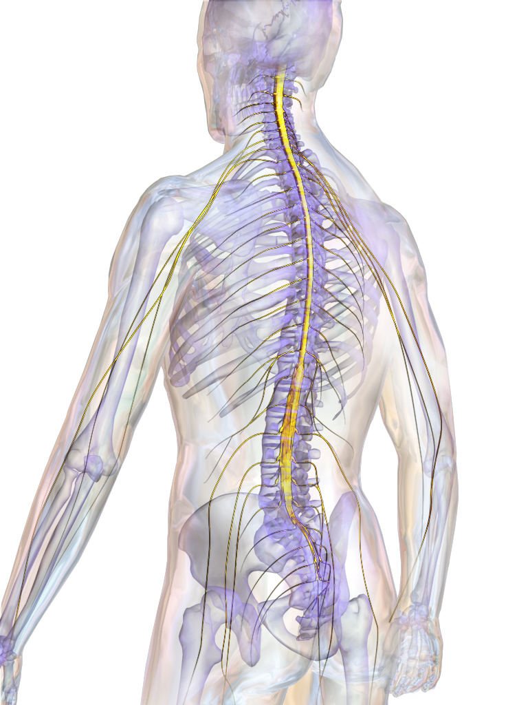
Structure of the Spinal Cord
The center of the spinal cord consists of gray matter, which is made up mainly of cell bodies of neurons, including interneurons and motor neurons. The gray matter is surrounded by white matter that consists mainly of myelinated axons of motor and sensory neurons. Spinal nerves, which connect the spinal cord to the PNS, exit from the spinal cord between vertebrae (see Figure 8.5.9).
Functions of the Spinal Cord
The spinal cord serves as an information superhighway. It passes messages from the body to the brain and from the brain to the body. Sensory (afferent) nerves carry nerve impulses to the brain from sensory receptor cells everywhere in and on the body. Motor (efferent) nerves carry nerve impulses away from the brain to glands, organs, or muscles throughout the body.
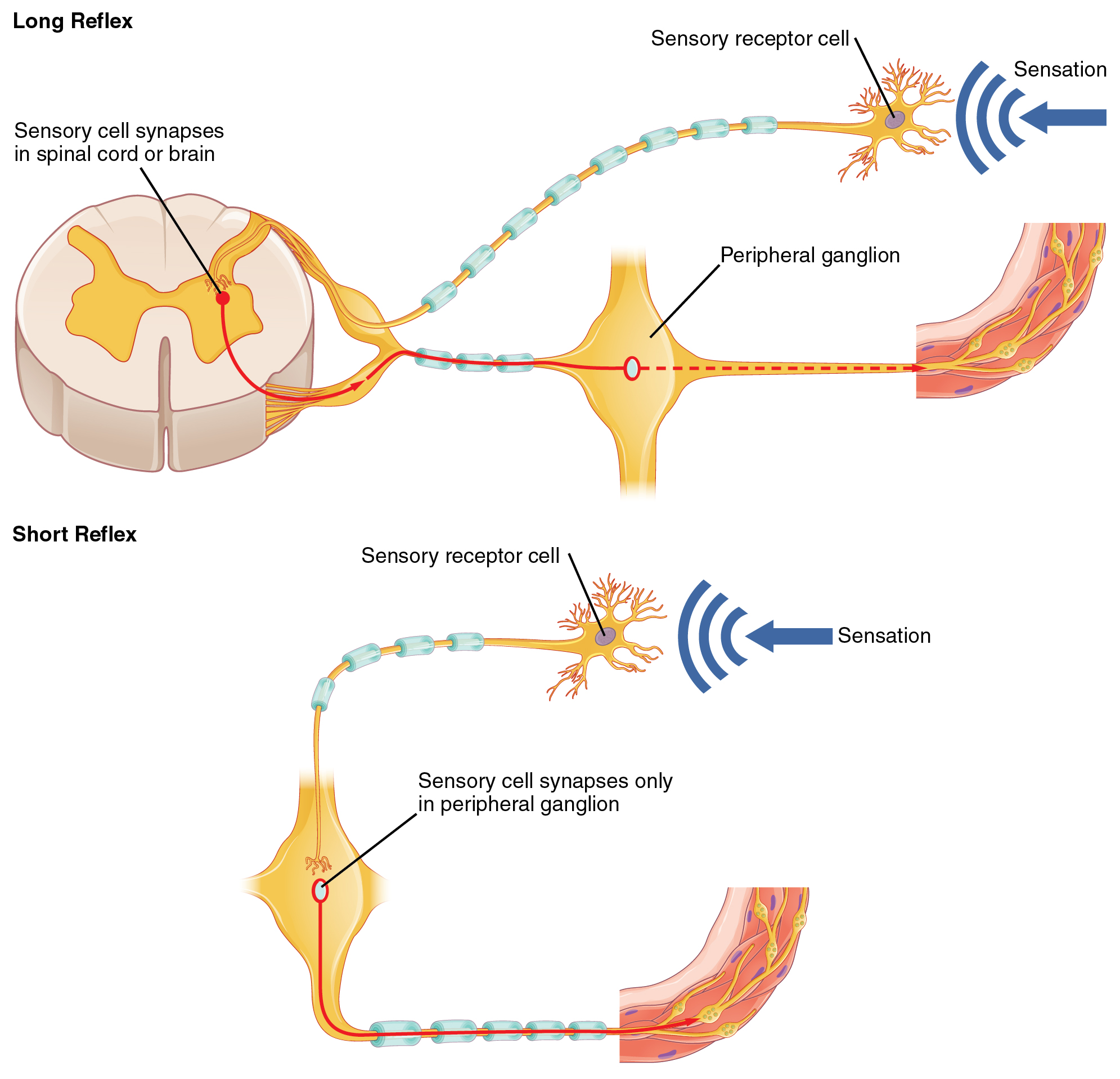
The spinal cord also independently controls certain rapid responses called reflexes without any input from the brain. You can see how this may happen in Figure 8.5.10. A sensory receptor responds to a sensation and sends a nerve impulse along a sensory nerve to the spinal cord. In the spinal cord, the message passes to an interneuron and from the interneuron to a motor nerve, which carries the impulse to a muscle. The muscle contracts in response. These neuron connections form a reflex arc, which requires no input from the brain. No doubt you have experienced such reflex actions yourself. For example, you may have reached out to touch a pot on the stove, not realizing that it was very hot. Virtually at the same moment that you feel the burning heat, you jerk your arm back and remove your hand from the pot.
Injuries to the Spinal Cord
Physical damage to the spinal cord may result in paralysis, which is loss of sensation and movement in part of the body. Paralysis generally affects all the areas of the body below the level of the injury, because nerve impulses are interrupted and can no longer travel back and forth between the brain and body beyond that point. If an injury to the spinal cord produces nothing more than swelling, the symptoms may be transient. However, if nerve fibres (axons) in the spinal cord are badly damaged, the loss of function may be permanent. Experimental studies have shown that spinal nerve fibres attempt to regrow, but tissue destruction usually produces scar tissue that cannot be penetrated by the regrowing nerves, as well other factors that inhibit nerve fibre regrowth in the central nervous system.
Feature: My Human Body
Each year, many millions of people have a stroke, and stroke is the second leading cause of death in adults. Stroke, also known as cerebrovascular accident, occurs when poor blood flow to the brain results in the death of brain cells. There are two main types of strokes:
- Ischemic strokes occur due to lack of blood flow because of a blood clot in an artery going to the brain.
- Hemorrhagic strokes occur due to bleeding from a broken blood vessel in the brain.
Either type of stroke may result in paralysis, loss of the ability to speak or comprehend speech, loss of bladder control, personality changes, and many other potential effects, depending on the part of the brain that is injured. The effects of a stroke may be mild and transient or more severe and permanent. A stroke may even be fatal. It generally depends on the type of stroke and how extensive it is.
Are you at risk of a stroke? The main risk factor for stroke is age — about two-thirds of strokes occur in people over the age of 65. There is nothing you can do about your age, but most other stroke risk factors can be reduced with lifestyle changes or medications. The risk factors include high blood pressure, tobacco smoking, obesity, high blood cholesterol, diabetes mellitus, and atrial fibrillation.
Chances are good that you or someone you know is at risk of a stroke, so it is important to recognize a stroke if one occurs. A stroke is a medical emergency, and the more quickly treatment is given, the better the outcome is likely to be. In the case of ischemic strokes, the use of clot-busting drugs may prevent permanent brain damage if administered within three or four hours of the stroke. Remembering the signs of a stroke is easy. They are summed up by the acronym FAST.
8.5 Summary
- The central nervous system is the part of the nervous system that includes the brain and spinal cord. It is physically protected by bones, meninges, and cerebrospinal fluid. It is chemically protected by the blood-brain barrier.
- The brain is the control center of the nervous system and of the entire organism. The brain uses a relatively large proportion of the body’s energy, primarily in the form of glucose.
- The brain is divided into three major parts, each with different functions: the hindbrain, the midbrain and the forebrain.
- Hindbrain consists of the medulla oblongata, the pons and the cerebellum, each of which perform specific functions.
- The midbrain is primarily responsible for motor movement and in auditory and visual processing.
- The forebrain contains many structures including:
- The cerebrum, which is further divided into left and right hemispheres. Each hemisphere has four lobes: frontal, parietal, temporal, and occipital. Each lobe is associated with specific senses or other functions.
- The cerebrum has a thin outer layer called the cerebral cortex. Its many folds give it a large surface area. This is where most information processing takes place.
- Inner structures of the brain include the hypothalamus — which controls the endocrine system via the pituitary gland — and the thalamus, which has several involuntary functions.
- The hippocampus and amygdala, which are part of the limbic system and play important roles in memory, learning and emotions.
- The spinal cord is a tubular bundle of nervous tissues that extends from the head down the middle of the back to the pelvis. It functions mainly to connect the brain with the peripheral nervous system. It also controls certain rapid responses called reflexes without any input from the brain.
- A spinal cord injury may lead to paralysis (loss of sensation and movement) of the body below the level of the injury, because nerve impulses can no longer travel up and down the spinal cord beyond that point.
8.5 Review Questions
- What is the central nervous system?
- How is the central nervous system protected?
- What is the overall function of the brain?
- Identify the three main parts of the brain and one function of each part.
- Describe the hemispheres of the brain.
- Explain and give examples of lateralization of the brain.
- Identify one function of each of the four lobes of the cerebrum.
-
-
- Summarize the structure and function of the cerebral cortex. Explain how the hypothalamus controls the endocrine system.
- Describe the spinal cord.
- What is the main function of the spinal cord?
- Explain how reflex actions occur.
- Why do severe spinal cord injuries usually cause paralysis?
- What do you think are some possible consequences of severe damage to the brain stem? How might this compare to the consequences of severe damage to the frontal lobe? Explain your answer.
- Information travels very quickly in the nervous system, but generally, the longer the path between areas, the longer it takes. Based on this, explain why you think reflexes often occur at the spinal cord level, and do not require input from the brain.
8.5 Explore More
What if we could look inside human brains? – Moran Cerf, TED-Ed, 2013.
The left brain vs. right brain myth – Elizabeth Waters, TED-Ed, 2017.
Split-brain patient ‘Joe’ being tested with stimuli presented in different visual fields,
markmcdermott, 2010.
Attributes
Figure 8.5.1
Sensory_Homunculus-en.svg by Popadius on Wikimedia Commons is used under a CC BY 3.0 (https://creativecommons.org/licenses/by/3.0/deed.en) license. (This is a derivative work from File:1421 Sensory Homunculus.jpg by OpenStax College)
Figure 8.5.2
Overview_of_Nervous_System by OpenStax on Wikimedia Commons is used under a CC BY 4.0 (https://creativecommons.org/licenses/by/4.0/deed.en) license.
Figure 8.5.3
Diagram_showing_the_brain_stem_which_includes_the_medulla_oblongata,_the_pons_and_the_midbrain_(2)_CRUK_294.svg by Cancer Research UK on Wikimedia Commons is used under a CC BY-SA 4.0 (https://creativecommons.org/licenses/by-sa/4.0) license.
Figure 8.5.4
Brain_bulbar_region.svg by Fvasconcellos on Wikimedia Commons is used under a CC BY 2.5 license (derivative work from Brain human sagittal section.svg by Patrick J. Lynch; Brain bulbar region.PNG by DO11.10).
Figure 8.5.5
Midbrain by BruceBlaus on Wikimedia Commons is used under a CC BY 3.0 (https://creativecommons.org/licenses/by/3.0) license.
Figure 8.5.6
BrainLobes by BruceBlaus on Wikimedia Commons is used under a CC BY 3.0 (https://creativecommons.org/licenses/by/3.0) license.
Figure 8.5.7
Diencephalon by OpenStax Wikimedia Commons is used under a CC BY 4.0 (https://creativecommons.org/licenses/by/4.0) license.
Figure 8.5.8
SpinalCord by BruceBlaus on Wikimedia Commons is used under a CC BY 3.0 (https://creativecommons.org/licenses/by/3.0) license.
Figure 8.5.9
Spinal_readjustment_3 by Tomwsulcer on Wikimedia Commons is used under a CC0 1.0 Universal Public Domain Dedication license (https://creativecommons.org/publicdomain/zero/1.0/deed.en).
Figure 8.5.10
1507_Short_and_Long_Reflexes by OpenStax on Wikimedia Commons is used under a CC BY 4.0 (https://creativecommons.org/licenses/by/4.0/deed.en) license.
References
Betts, J. G., Young, K.A., Wise, J.A., Johnson, E., Poe, B., Kruse, D.H., Korol, O., Johnson, J.E., Womble, M., DeSaix, P. (2013, 28 May). Figure 14.23 The sensory homunculus [digital image]. In Anatomy and Physiology (Section 14.2). OpenStax. https://openstax.org/books/anatomy-and-physiology/pages/14-2-central-processing
Betts, J. G., Young, K.A., Wise, J.A., Johnson, E., Poe, B., Kruse, D.H., Korol, O., Johnson, J.E., Womble, M., DeSaix, P. (2013, 28 May). Figure 15.8 Short and long reflexes [digital image]. In Anatomy and Physiology (Section 15.2). OpenStax. https://openstax.org/books/anatomy-and-physiology/pages/15-2-autonomic-reflexes-and-homeostasis
Betts, J. G., Young, K.A., Wise, J.A., Johnson, E., Poe, B., Kruse, D.H., Korol, O., Johnson, J.E., Womble, M., DeSaix, P. (2016, May 18). Figure 12.2 Central and peripheral nervous system [digital image]. In Anatomy and Physiology (Section 12.1). https://openstax.org/books/anatomy-and-physiology/pages/12-1-basic-structure-and-function-of-the-nervous-system
Betts, J. G., Young, K.A., Wise, J.A., Johnson, E., Poe, B., Kruse, D.H., Korol, O., Johnson, J.E., Womble, M., DeSaix, P. (2016, May 18). Figure 13.11 The diencephalon [digital image]. In Anatomy and Physiology (Section 13.2). https://openstax.org/books/anatomy-and-physiology/pages/13-2-the-central-nervous-system
Blausen.com staff. (2014). Medical gallery of Blausen Medical 2014. WikiJournal of Medicine, 1 (2). DOI:10.15347/wjm/2014.010. ISSN 2002-4436
markmcdermott. (2010). Split-brain patient ‘Joe’ being tested with stimuli presented in different visual fields. YouTube. https://www.youtube.com/user/markmcdermott/search?query=split
Mayo Clinic Staff. (n.d.). Stroke [online article]. MayoClinic.org. https://www.mayoclinic.org/diseases-conditions/stroke/symptoms-causes/syc-20350113
Soton Brain Hub. (2016, July 29). The limbic system. YouTube. https://www.youtube.com/watch?v=jcrWPo_s6EE&feature=youtu.be
TED-Ed. (2013, January 31). What if we could look inside human brains? – Moran Cerf. YouTube. https://www.youtube.com/watch?v=sewhbmh0ECg&feature=youtu.be
TED-Ed. (2017, July 24). The left brain vs. right brain myth – Elizabeth Waters. YouTube. https://www.youtube.com/watch?v=ZMSbDwpIyF4&feature=youtu.be
One of two main divisions of the nervous system that includes the brain and spinal cord.
The central nervous system organ inside the skull that is the control center of the nervous system.
A thin, tubular bundle of central nervous system tissue that extends from the brainstem down the back to the pelvis and connects the brain with the peripheral nervous system.
One of two major divisions of the nervous system that consists of all the nervous tissue that lies outside the central nervous system.
A three-layered membrane that encloses and protects the brain and spinal cord and contains cerebrospinal fluid.
Clear fluid produced by the brain that forms a thin layer within the meninges and provides protection and cushioning for the brain and spinal cord.
A highly selective membrane formed of epithelial cells that separates circulating blood from extracellular fluid in the brain and spinal cord.
A functional unit of the nervous system that transmits nerve impulses; also called a nerve cell.
A class of nervous system cell that provides support for neurons and helps them transmit nerve impulses.
Glucose (also called dextrose) is a simple sugar with the molecular formula C6H12O6. Glucose is the most abundant monosaccharide, a subcategory of carbohydrates. Glucose is mainly made by plants and most algae during photosynthesis from water and carbon dioxide, using energy from sunlight.
A multi-branched polysaccharide of glucose that serves as a form of energy storage in animals, fungi, and bacteria.
One of the three major regions of the human brain, located at the lower back part of the brain. It includes most of the brainstem and a dense coral-shaped structure called the cerebellum. The brainstem is one of the most important parts of the entire central nervous system, because it connects the brain to the spinal cord and coordinates many vital functions, such as breathing and heartbeat.
The part of the brain below the cerebrum and behind the brain stem that coordinates body movements.
A long stem-like structure which makes up part of the brainstem. It is anterior and partially inferior to the cerebellum. It is responsible for autonomic (involuntary) functions ranging from vomiting to sneezing.
Part of the central nervous system, located at the base of the brain, between the medulla oblongata and the midbrain. It is part of the brainstem. The pons serves as a message station between several areas of the brain. It helps relay messages from the cortex and the cerebellum
A diffuse network of nerve pathways in the brainstem connecting the spinal cord, cerebrum, and cerebellum, and mediating the overall level of consciousness.
The largest part of the brain that controls conscious functions such as reasoning and sight.
One of two halves (left and right) of the cerebrum of the human brain.
A thick band of nerve fibers that divides the cerebral cortex lobes into left and right hemispheres. It connects the left and right sides of the brain, allowing for communication between both hemispheres.
The concentration of particular functions in one hemisphere of the cerebrum of the brain.
A part of each hemisphere of the cerebrum that controls executive functions such as reasoning and language.
The part of each hemisphere of the cerebrum that is involved in functions such as touch, reading, and arithmetic.
Part of each hemisphere of the cerebrum that is involved in functions such as hearing, memories, and sensory integration.
A part of each hemisphere of the cerebrum that is dedicated almost solely to vision.
The highly folded, thin outer layer of the cerebrum where most information processing in the brain takes place.
A set of brain structures located on both sides of the thalamus, immediately beneath the medial temporal lobe of the cerebrum primarily in the forebrain. It supports a variety of functions including emotion, behavior, motivation, long-term memory, and olfaction. Emotional life is largely housed in the limbic system, and it critically aids the formation of memories.
A part of the brain that secretes hormones and connects the brain with the endocrine system.
division of the peripheral nervous system that controls involuntary activities
An endocrine hormone secreted by the pituitary gland that controls a variety of functions, including during childbirth to stimulate uterine contractions and during lactation to trigger milk letdown.
A hormone made by the hypothalamus in the brain and stored in the posterior pituitary gland. It tells your kidneys how much water to conserve. ADH constantly regulates and balances the amount of water in your blood. Higher water concentration increases the volume and pressure of your blood.
The master gland of the endocrine system that secretes many hormones, the majority of which regulate other endocrine glands.
The inner part of the brain that is a major hub for nerve impulses traveling back and forth between the cerebrum and spinal cord.
A complex brain structure embedded deep into temporal lobe. It has a major role in learning and memory.
The loss of sensation and movement in part of the body, such as may occur with a stroke or spinal cord injury.
A cerebrovascular accident in which a broken artery or blood clot results in lack of blood flow to part of the brain, causing death of brain cells.

_CRUK_294.svg)