10.8 Case Study Conclusion: Wearing His Heart on His Sleeve
Created by CK-12 Foundation/Adapted by Christine Miller
Case Study Conclusion: Wearing His Heart on His Sleeve
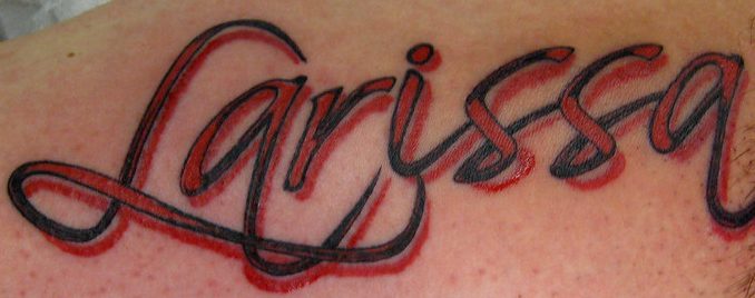
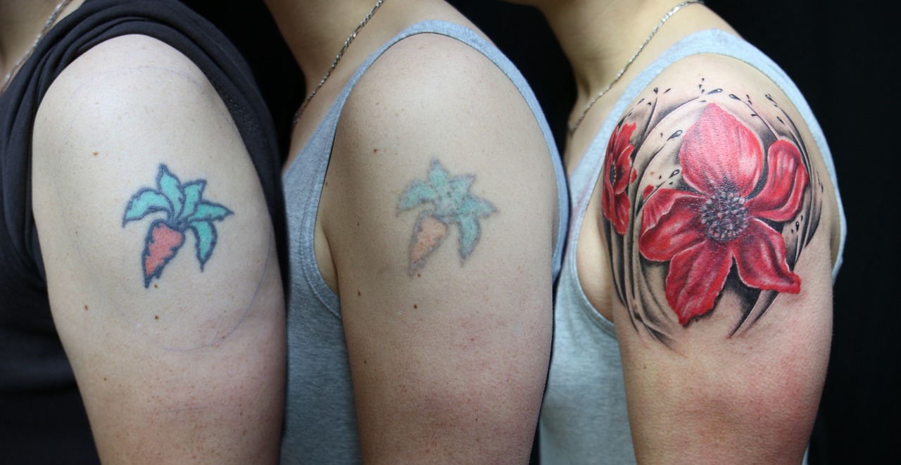
Are you still wondering whether Ayko, who you read about in the beginning of this chapter, actually got a tattoo of his new girlfriend’s name on his arm? Figure 10.8.1 is your answer! Let’s hope his love for Larissa — and for the artwork — lasts as long as his tattoo. According to a poll conducted for Global TV by Ipsos Reid in 2012, 10% of Canadian and 11% of American adults regret getting a tattoo. Although laser tattoo removal is available, it does not always work fully, can cause pain and scarring, and is expensive and time-consuming. Some people who regret a tattoo opt instead (or additionally) to cover it with another tattoo, see Figure 10.8.2 below.
Why are tattoos essentially permanent? Tattoos are created by inserting a needle containing pigment through the epidermis and into the dermis of the skin. The pigment is injected into the dermal layer, creating the design. The pigment can remain in the dermal layer for a person’s lifetime for a few reasons. One, unlike the thinner outer epidermal layer, the dermis is not continually shed and replaced, so the pigment generally stays put. Two, the pigments used in tattooing mainly consist of large particles. When you get a tattoo, the penetration of the skin and insertion of foreign particles causes an immune response in which white blood cells attempt to engulf and remove the pigment. Because most of the pigment particles are so large, however, they cannot be removed from the dermis by the immune cells, and the design remains.
In laser tattoo removal, pulses from a high-intensity laser are applied to the tattoo and absorbed by the pigments. This breaks up the large pigment particles into particles that are small enough to be removed by the immune system. The pigments may then be excreted out of the body, or moved to other areas of the body, such as the lymph nodes. Different wavelengths of laser energy are often required to remove different colours of pigments, because they absorb different wavelengths of light. Generally, blue and black are the easiest colours to remove. Green, red, and yellow tend to be the hardest to remove. It may take as many as six to ten laser treatments — with a few weeks of recovery time in between — to remove a tattoo. Some tattoos can never be completely removed.
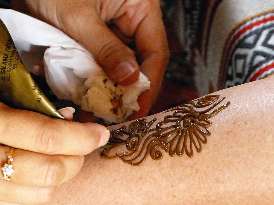
Why are mehndi designs (like Ayko’s trial “henna tattoo”) not permanent? Unlike real tattoos, henna paste is applied on the surface of the skin (shown below in Figure 10.8.3), and not injected into the skin with a needle. The dye molecules simply migrate from the paste into the top layer of the epidermis, the stratum corneum.
As you have learned, the stratum corneum consists of dead, keratin-filled keratinocytes, which are continually shed and replaced with new cells from the layers below. As a result, mehndi is not permanent. The design is lost as the cells that contain the dye are shed and replaced.
As you read in the beginning of this chapter, mehndi is often applied to the palms of the hands and soles of the feet, which generally results in a darker stain than other areas of the body. This is because the stratum corneum is thicker in these regions, so the dye penetrates through more layers of cells, making the design appear darker. What else is different about the epidermis of the palms and soles? You may recall that these regions are the only place where there is a fifth layer of epidermis — the stratum lucidum — making the skin in these areas even thicker and tougher.
Hopefully, Ayko thought carefully about the potential emotional and social implications of getting a tattoo — and learned how difficult they are to remove — before getting a real one. Health and safety should also be of utmost concern to anyone considering getting a tattoo. As you have learned in this chapter, the skin acts as a barrier against dangerous pathogens and substances. When you penetrate the skin using a needle, it can introduce harmful viruses and bacteria directly into the dermis, where the blood vessels are. Tattoo artists and shops need to take precautions to protect their clients against diseases that can be transmitted through blood (such as HIV and hepatitis), as well as bacterial infections. The tattoo artist should wear disposable gloves and a mask, use new and unopened needles and ink tubes, and properly sterilize other equipment. Even if the artist takes all the proper precautions, there is still a chance that the unopened ink could have been contaminated with pathogens during the production process. The shop should be aware of any ink recalls. Anyone getting a tattoo should make sure their artist and shop strictly adhere to all local health and safety regulations.
The risk of disease is not the only risk from tattoos. The pigments in tattoos may contain heavy metals and other potentially toxic substances. Tattoo parlours are regulated by provincial guidelines in Canada, and these guidelines vary from province to province — but these guidelines are mainly concerned with sterilization of equipment and don’t address anything about pigments. A recent study published in the scientific journal Nature (Scientific Reports) showed that pigments from tattoos may migrate from a person’s tattoos into their lymph nodes. Among the substances that make up the tattoo ink that migrated were aluminum, chromium, iron, nickel and copper – all considered “toxic”.
Additionally, people can sometimes have an allergic reaction to the pigments, or develop scarring or granulomas (small bumps of tissue due to an immune response) around the tattoo. Rarely, people can experience temporary swelling or burning of their tattoos when they get scanned in an MRI machine for a medical procedure. Clearly, people should think carefully about the potential health implications before getting a tattoo.
Fortunately, Ayko found a reputable and safe tattoo artist, and is not experiencing any ill effects from his tattoo. He is happy with his tattoo, at least for now. Tattoos — and other kinds of decoration of the integumentary system — are forms of artistic, personal, and cultural expression that have been used by many cultures over the course of human history. The system that protects us from the elements, helps us maintain homeostasis, and mediates our interactions with the outside world also happens to be easily modifiable! Whether it is a haircut, makeup, beard style, nail polish, piercing or a tattoo, humans have a variety of ways of altering our integumentary system, which changes our outward appearance and what we communicate to others.
Chapter 10 Summary
In this chapter, you learned about the structures and functions of the organs of the integumentary system. Specifically, you learned that:
- The integumentary system consists of the skin, hair, and nails. Functions of the integumentary system include providing a protective covering for the body, sensing the environment, and helping the body maintain homeostasis.
- The skin’s main functions include preventing water loss from the body, serving as a barrier to the entry of microorganisms, synthesizing vitamin D, blocking UV light, and helping to regulate body temperature.
- The skin consists of two distinct layers: a thinner outer layer called the epidermis, and a thicker inner layer called the dermis.
-
- The epidermis consists mainly of epithelial cells called keratinocytes, which produce keratin. New keratinocytes form at the bottom of the epidermis. They become filled with keratin and die as they move upward toward the surface of the skin, where they form a protective, waterproof layer.
- The dermis consists mainly of tough connective tissues that provide strength and stretch, as well as almost all skin structures, including blood vessels, sensory receptors, hair follicles, and oil and sweat glands.
- Cell types in the epidermis include keratinocytes (which make up 90 per cent of epidermal cells), melanocytes that produce melanin, Langerhans cells that fight pathogens in the skin, and Merkel cells that respond to light touch.
- In most parts of the body, the epidermis consists of four distinct layers. A fifth layer occurs only in the epidermis of the palms of the hands and soles of the feet.
-
- The innermost layer of the epidermis is the stratum basale, which contains stem cells that divide to form new keratinocytes.
- The next layer is the stratum spinosum, which is the thickest layer, and contains Langerhans cells and spiny keratinocytes.
- This is followed by the stratum granulosum, in which keratinocytes are filling with keratin and beginning to die.
- The stratum lucidum is next, but only on the palms and soles. It consists of translucent dead keratinocytes.
- The outermost layer is the stratum corneum, which consists of flat, dead, tightly packed keratinocytes that form a tough, waterproof barrier for the rest of the epidermis.
- The epidermis protects underlying tissues from physical damage and pathogens. Melanin in the epidermis absorbs and protects underlying tissues from UV light. The epidermis also prevents loss of water from the body and synthesizes vitamin D.
-
- Melanin is the main pigment that determines the colour of human skin. However, the pigments carotene and hemoglobin also contribute to skin colour, especially in skin with low levels of melanin.
- The surface of healthy skin normally is covered by vast numbers of bacteria representing about one thousand species from 19 phyla. Different areas of the body provide diverse habitats for skin microorganisms. Usually, microorganisms on the skin keep each other in check unless their balance is disturbed.
- The thicker inner layer of the skin — the dermis — has two layers. The upper papillary layer has papillae extending upward into the epidermis and loose connective tissues. The lower reticular layer has denser connective tissues and structures, such as glands and hair follicles. Glands in the dermis include eccrine and apocrine sweat glands, as well as sebaceous glands. Hair follicles are structures where hairs originate.
- Functions of the dermis include cushioning subcutaneous tissues, regulating body temperature, sensing the environment, and excreting wastes. The dense connective tissues of the dermis provide cushioning. The dermis regulates body temperature mainly by sweating and by vasodilation or vasoconstriction. The many tactile sensory receptors in the dermis make it the main organ for the sense of touch. Wastes excreted in sweat include excess water, electrolytes, and certain metabolic wastes.
- Hair is a filament that grows from a hair follicle in the dermis of the skin. It consists mainly of tightly packed, dead keratinocytes that are filled with keratin. The human body is almost completely covered with hair follicles.
- Hair helps prevent heat loss from the head and protects its skin from UV light. Hair in the nose filters incoming air, and the eyelashes and eyebrows keep harmful substances out of the eyes. Hair all over the body provides tactile sensory input. The eyebrows also play a role in nonverbal communication.
- The part of a hair that is within the follicle is the hair root. This is the only living part of a hair. The part of a hair that is visible above the skin surface is the hair shaft. It consists of dead cells.
-
- Hair growth begins inside a follicle when stem cells within the follicle divide to produce new keratinocytes.
- A hair shaft has three zones: the outermost zone called the cuticle, the middle zone called the cortex, and the innermost zone called the medulla.
- Genetically controlled, visible characteristics of hair include hair colour, hair texture, and the extent of balding in adult males. Melanin (eumelanin and/or pheomelanin) is the pigment that gives hair its colour. Aspects of hair texture include curl pattern, thickness, and consistency.
- Among mammals, humans are nearly unique in having undergone significant loss of body hair during their evolution, probably because sweat evaporates more quickly from less hairy skin. Curly hair also is thought to have evolved at some point during human evolution, perhaps because it provided better protection from UV light.
- Hair has social significance for human beings, being an indicator of biological sex, age, and ethnic ancestry. Human hair also has cultural significance. For example, hairstyle may be an indicator of social group membership.
- Nails consist of sheets of dead, keratin-filled keratinocytes. The keratin in nails makes them hard but flexible. They help protect the ends of the fingers and toes, enhance the sense of touch in the fingertips, and may be used as tools.
- A nail has three main parts: the nail root, which is under the epidermis; the nail plate, which is the visible part of the nail; and the free margin, which is the distal edge of the nail. Other structures under or around a nail include the nail bed, cuticle, and nail fold.
- A nail grows from a deep layer of living epidermal tissues, called the nail matrix, at the proximal end of the nail. Stem cells in the nail matrix keep dividing to allow nail growth, forming first the nail root and then the nail plate as the nail continues to grow longer and emerges from the epidermis.
- Fingernails grow faster than toenails. Actual rates of growth depend on many factors, such as age, sex, and season.
- The colour of the nail bed can be used to quickly assess oxygen and blood flow in a patient. How the nail plate grows out can reflect recent health problems, such as illness or nutrient deficiency. Nails — and especially toenails — are prone to fungus infections. Nails are more permeable than skin and can absorb several harmful substances, such as herbicides.
- Skin cancer is a disease in which skin cells grow out of control. It is caused mainly by excessive exposure to UV light, which damages DNA.
- There are three common types of skin cancer: basal cell carcinoma, squamous cell carcinoma, and melanoma. Carcinomas are more common and unlikely to metastasize. Melanoma is rare and likely to metastasize. It causes most skin cancer deaths.
- Besides exposure to UV light, risk factors for skin cancer include having light coloured skin, having many moles, and a family history of skin cancer, among several others.
Now that you have learned about the organs on surface of the body, read the next chapter to travel inside and learn about the skeletal system, which protects and supports us internally, among other functions.
Chapter 10 Review
- Describe one way in which the integumentary system works with another organ system to carry out a particular function.
-
- Describe two types of waterproofing used in the integumentary system. Include the types of molecules and where they are located.
- Explain why nails enhance touch sensations.
- Why do you think light coloured skin is a risk factor for skin cancer?
- Describe the similarities between how the epidermis, hair, and nails all grow.
- What does the whitish crescent-shaped area at the base of your nails (toward your hands) represent? What is its function?
- What is one difference between human hair and the hair of non-human primates?
- Describe the relationship between skin and hair.
- What kind of skin cancer is a cancer of a type of stem cell?
- For the skin and hair, describe one way in which they each protect the body against pathogens.
- If sweat glands are in the dermis, how is sweat released to the surface of the body?
- Explain why you think that physicians usually insist that patients remove any nail polish before having surgery.
- Describe generally how the brain gets touch information from the skin.
Attributions
Figure 10.8.1
Larissa Tattoo4039922685_46bf0bcfe5_c by Micael Faccio on Flickr is used under a CC BY 2.0 (https://creativecommons.org/licenses/by/2.0/) license.
Figure 10.8.2
Tattoo laser and cover-631211_1280 [photo] by Herco Roelofs on Pixabay is used under the Pixabay License (https://pixabay.com/ja/service/license/).
Figure 10.8.3
henna-tattoo-abu-dhabi by MarieFrance on Pixabay is used under the Pixabay License (https://pixabay.com/ja/service/license/).
References
Global News Staff. (2017, September 15). Health: ‘Toxic’ tattoo ink particles can travel to your lymph nodes: study. Globalnews.ca. https://globalnews.ca/news/3746925/tattoo-ink-safety-lymph-nodes/
Ipsos Reid. (2012). “Two in ten Canadians (22%), Americans (21%)
have a tattoo; One in ten tattooed Canadians (10%), Americans (11%) regret it” [News release]. Ipsos.com. https://www.ipsos.com/sites/default/files/publication/2012-01/5490.pdf
Rideout, K. (2010, July). Comparison of guidelines and regulatory frameworks for personal services establishments. National Collaborating Centre for Environmental Health. https://www.ncceh.ca/sites/default/files/PSE_Guidelines_Comparison_Table_July%202010.pdf
Schreiver, I., Hesse, B., Seim, C. et al. Synchrotron-based ν-XRF mapping and μ-FTIR microscopy enable to look into the fate and effects of tattoo pigments in human skin. Scientific Reports 7,11395. https://doi.org/10.1038/s41598-017-11721-z
The outer layer of skin that consists mainly of epithelial cells and lacks nerve endings, blood vessels, and other structures.
The inner layer of skin that is made of tough connective tissue and contains blood vessels, nerve endings, hair follicles, and glands.
The outer layer of the skin (epidermis). It serves as the primary barrier between the body and the environment.
A tough, fibrous protein in skin, hair, and nails.
A type of epithelial cell found in the skin, hair, and nails that produces keratin.
a thin, clear layer of dead skin cells in the epidermis named for its translucent appearance under a microscope. It is readily visible by light microscopy only in areas of thick skin, which are found on the palms of the hands and the soles of the feet.
A microorganism which causes disease.
The body system comprised of skin and its appendages acting to protect the body from various kinds of damage, such as loss of water or damages from outside.
The ability of an organism to maintain constant internal conditions despite external changes.
The major organ of the integumentary system that covers and protects the body and helps maintain homeostasis, for example, by regulating body temperature.
A filament made of tightly packed, keratin-filled keratinocytes that grows out of a hair follicle in the dermis of the skin.
accessory organ of the skin made of sheets of dead keratinocytes at the distal ends of the fingers and toes
A form of electromagnetic radiation with wavelength shorter than that of visible light but longer than X-rays. UV radiation is present in sunlight, and constitutes about 10% of the total electromagnetic radiation output from the sun.
One of the four basic types of tissue, connective tissue is found in between other tissues everywhere in the body, including the nervous system and generally forms a framework and support structure for body tissues and organs.
Specialized nerve cell that responds to a particular type of stimulus such as light or chemicals by generating a nerve impulse.
A structure in the dermis of skin where a hair originates.
An exocrine gland in the dermis of the skin that produces the salty fluid called sweat through a duct to the skin surface.
A special skin cell that is responsible for producing melanin.
A brown pigment produced by melanocytes in the skin that gives skin most of its color and prevents UV light from penetrating the skin.
A cell found in the epidermis that functions as an antigen-presenting cell which binds antigens entering through the skin.
Oval-shaped mechanoreceptors essential for light touch sensation and found in the skin.
The innermost (deepest) layer of the epidermis. Consists of a single layer of columnar or cuboidal basal cells.
A layer of the epidermis found between the stratum granulosum and stratum basale. The main function of the stratum spinosum is to allow keratinocytes (cells that produce keratin) to mature.
A thin layer of cells in the epidermis. Keratinocytes migrating from the underlying stratum spinosum become known as granular cells in this layer. Function is to help to form a waterproof barrier that functions to prevent fluid loss from the body.
A pigment in the epidermis that gives skin a yellowish tint, especially in skin with low levels of melanin.
An oxygen-binding protein containing iron that is the principal component of red blood cells.
Any member of a large group of unicellular microorganisms which have cell walls but lack organelles and an organized nucleus, including some which can cause disease.
The upper layer of the dermis with papillae extending upward into the epidermis.
The lower layer of the dermis that gives the dermis strength and elasticity and contains many dermal structures such as glands and hair follicles.
The major sweat glands of the human body, sometimes called merocrine glands, found in virtually all skin, with the highest density in palm and soles, then on the head, but much less on the trunk and the extremities.
Sweat glands that secrete their products into a hair follicle. Present only in certain places in the human body including the armpits, nipples, ear canal, eyelids, and parts of the external genitalia.
A gland in the dermis of the skin that produces sebum, an oily substance that waterproofs the skin and hair.
The widening of blood vessels. It results from relaxation of smooth muscle cells within the vessel walls, in particular in the large veins, large arteries, and smaller arterioles. The process is the opposite of vasoconstriction, which is the narrowing of blood vessels.
A narrowing of blood vessels so less blood can flow through them.
The part of a hair that is located within the hair follicle and consists of living keratinocytes.
A part of a hair that is visible above the surface of the skin and consists of dead keratinocytes.
The outermost part of the hair shaft. It is formed from dead cells, overlapping in layers, which form scales that strengthen and protect the hair shaft.
Located between the hair cuticle and medulla and is the thickest hair layer. It also contains most of the hair's pigment, giving the hair its color. The pigment in the cortex is melanin, which is also found in skin.
The innermost layer of the hair shaft. This nearly invisible layer is the most soft and fragile, and serves as the pith or marrow of the hair.
A dark pigment that predominates in black and brunette hair. There are two different types of eumelanin (brown eumelanin and black eumelanin). A small amount of brown eumelanin in the absence of other pigments causes blond hair.
A lighter pigment found in red hair, and is concentrated in the redder areas of the skin such as the lips. Because people with red hair are less able to make the dark eumelanin pigment, their skin is generally quite pale and burns easily with sun exposure.
A portion of a nail that is under the surface of the skin at the proximal end of the nail.
visible part of a nail that is external to the skin
The part of the nail that protrudes beyond the end of the finger or toe; the part that is cut or filed to keep the nail trimmed.
The pink skin under the nail plate visible through the nail.
A layer of clear skin located along the bottom edge of your finger or toe. The cuticle function is to protect new nails from bacteria when they grow out from the nail root. The area around the cuticle is delicate. It can get dry, damaged, and infected.
The tissue that encloses the nail matrix at the root of the nail.
A group of diseases involving abnormal cell growth with the potential to invade or spread to other parts of the body.
The most common type of skin cancer that occurs in basal cells of the epidermis and rarely metastasizes.
A common type of skin cancer that affects squamous cells in the epidermis and rarely metastasizes.
A rare but most serious type of skin cancer that affects melanocytes and usually metastasizes if not treated.
