10.2 Introduction to the Integumentary System
Created by CK-12 Foundation/Adapted by Christine Miller
Art for All Eras
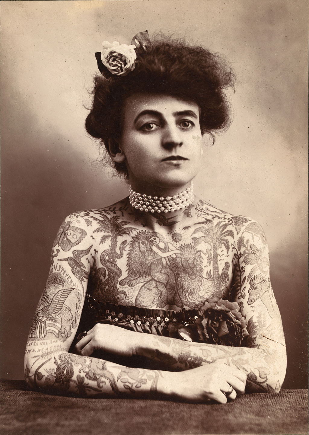
Pictured in Figure 10.2.1, is Maud Stevens Wagner, a tattoo artist from 1907. Tattoos are not just a late 20th and early 21st century trend. They have been popular in many eras and cultures. Tattoos literally illustrate the biggest organ of the human body: the skin. The skin is very thin, but it covers a large area — about 2 m2 in adults. The skin is the major organ in the integumentary system.
What Is the Integumentary System?
In addition to the skin, the integumentary system includes the hair and nails, which are organs that grow out of the skin. Because the organs of the integumentary system are mostly external to the body, you may think of them as little more than accessories, like clothing or jewelry, but they serve vital physiological functions. They provide a protective covering for the body, sense the environment, and help the body maintain homeostasis.
The Skin
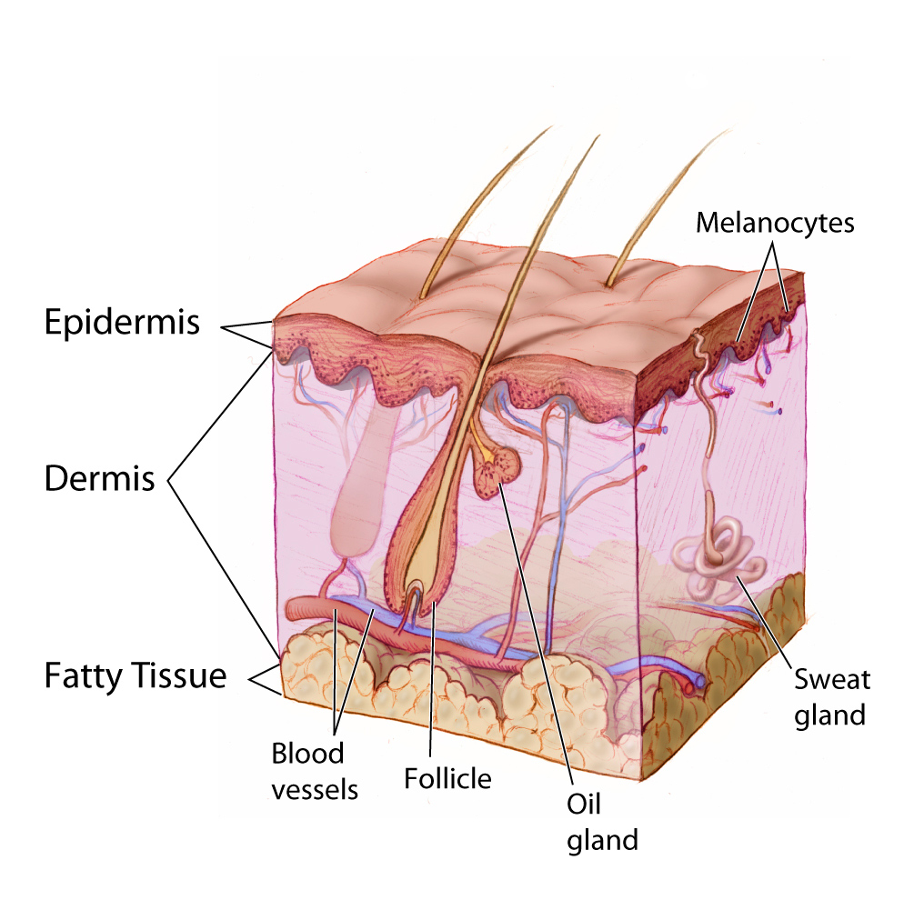
The skin is remarkable not only because it is the body’s largest organ: the average square inch of skin has 20 blood vessels, 650 sweat glands, and more than 1,000 nerve endings. Incredibly, it also has 60,000 pigment-producing cells. All of these structures are packed into a stack of cells that is just 2 mm thick. Although the skin is thin, it consists of two distinct layers: the epidermis and dermis, as shown in the diagram (Figure 10.2.2).
Outer Layer of Skin
The outer layer of skin is the epidermis. This layer is thinner than the inner layer (the dermis). The epidermis consists mainly of epithelial cells, called keratinocytes, which produce the tough, fibrous protein keratin. The innermost cells of the epidermis are stem cells that divide continuously to form new cells. The newly formed cells move up through the epidermis toward the skin surface, while producing more and more keratin. The cells become filled with keratin and die by the time they reach the surface, where they form a protective, waterproof layer. As the dead cells are shed from the surface of the skin, they are replaced by other cells that move up from below. The epidermis also contains melanocytes, the cells that produce the brown pigment melanin, which gives skin most of its colour. Although the epidermis contains some sensory receptor cells — called Merkel cells — it contains no nerves, blood vessels, or other structures.
Inner Layer of Skin
The dermis is the inner, thicker layer of skin. It consists mainly of tough connective tissue, and is attached to the epidermis by collagen fibres. The dermis contains many structures (as shown in Figure 10.2.2), including blood vessels, sweat glands, and hair follicles, which are structures where hairs originate. In addition, the dermis contains many sensory receptors, nerves, and oil glands.
Functions of the Skin
The skin has multiple roles in the body. Many of these roles are related to homeostasis. The skin’s main functions are preventing water loss from the body and serving as a barrier to the entry of microorganisms. Another function of the skin is synthesizing vitamin D, which occurs when the skin is exposed to ultraviolet (UV) light. Melanin in the epidermis blocks some of the UV light and protects the dermis from its damaging effects.
Another important function of the skin is helping to regulate body temperature. When the body is too warm, for example, the skin lowers body temperature by producing sweat, which cools the body when it evaporates. The skin also increases the amount of blood flowing near the body surface through vasodilation (widening of blood vessels), bringing heat from the body core to radiate out into the environment. The sweaty hair and flushed skin of the young man pictured in Figure 10.2.3 reflect these skin responses to overheating.

Hair
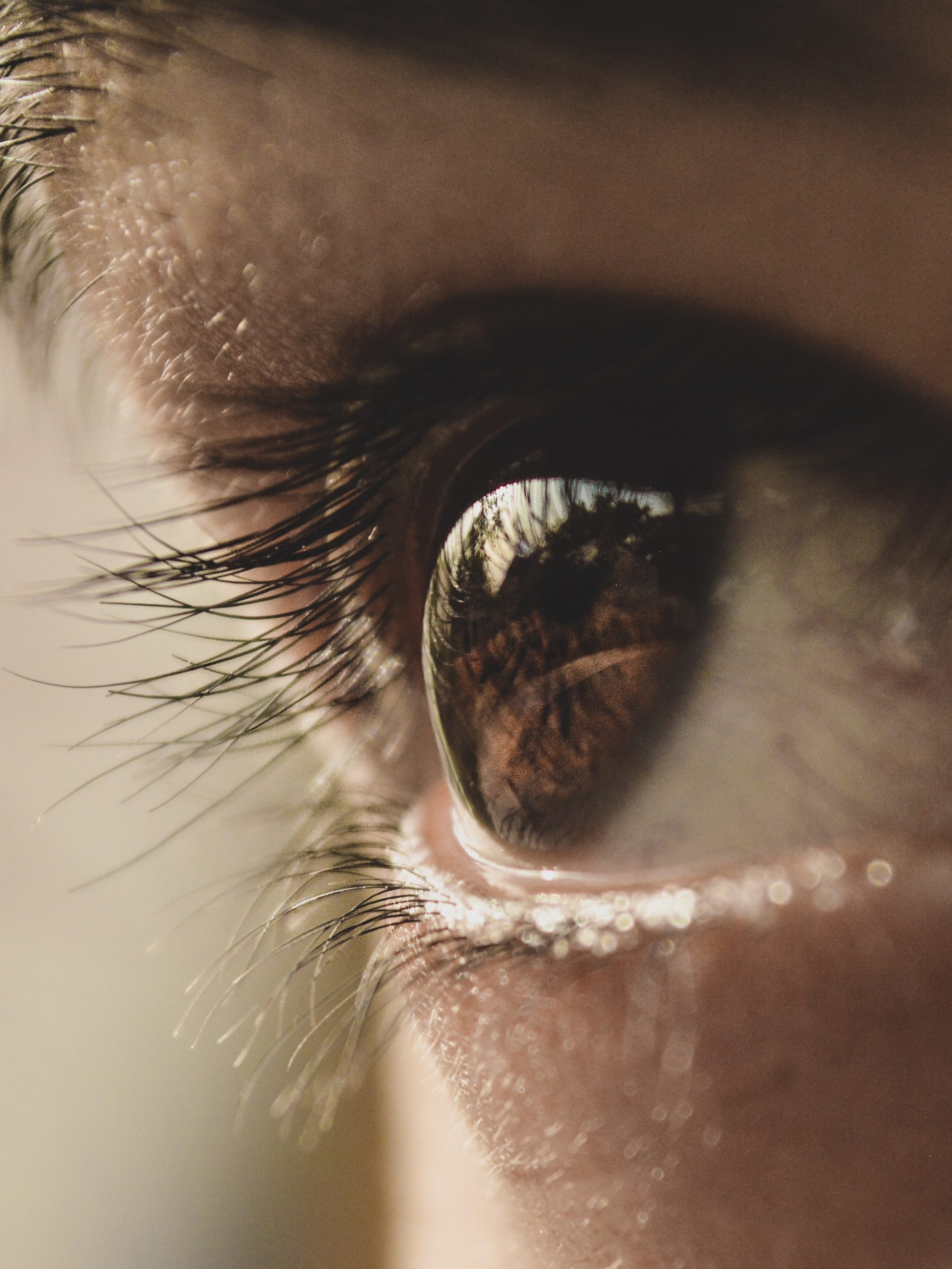
Hair is a fibre found only in mammals. It consists mainly of keratin-producing keratinocytes. Each hair grows out of a follicle in the dermis. By the time the hair reaches the surface, it consists mainly of dead cells filled with keratin. Hair serves several homeostatic functions. Head hair is important in preventing heat loss from the head and protecting its skin from UV radiation. Hairs in the nose trap dust particles and microorganisms in the air, and prevent them from reaching the lungs. Hair all over the body provides sensory input when objects brush against it, or when it sways in moving air. Eyelashes and eyebrows (see Figure 10.2.4) protect the eyes from water, dirt, and other irritants.
Nails
Fingernails and toenails consist of dead keratinocytes filled with keratin. The keratin makes them hard but flexible, which is important for the functions they serve. Nails prevent injury by forming protective plates over the ends of the fingers and toes. They also enhance sensation by acting as a counterforce to the sensitive fingertips when objects are handled. In addition, the fingernails can be used as tools.
Interactions with Other Organ Systems
The skin and other parts of the integumentary system work with other organ systems to maintain homeostasis.
- The skin works with the immune system to defend the body from pathogens by serving as a physical barrier to microorganisms.
- Vitamin D is needed by the digestive system to absorb calcium from food. By synthesizing vitamin D, the skin works with the digestive system to ensure that calcium can be absorbed.
- To control body temperature, the skin works with the cardiovascular system to either lose body heat, or to conserve it through vasodilation or vasoconstriction.
- To detect certain sensations from the outside world, the nervous system depends on nerve receptors in the skin.
10.2 Summary
- The integumentary system consists of the skin, hair, and nails. Functions of the integumentary system include providing a protective covering for the body, sensing the environment, and helping the body maintain homeostasis.
- The skin consists of two distinct layers: a thinner outer layer called the epidermis, and a thicker inner layer called the dermis.
- The epidermis consists mainly of epithelial cells called keratinocytes, which produce keratin. New keratinocytes form at the bottom of the epidermis. They become filled with keratin and die as they move upward toward the surface of the skin, where they form a protective, waterproof layer.
- The dermis consists mainly of tough connective tissues and many structures, including blood vessels, sensory receptors, nerves, hair follicles, and oil and sweat glands.
- The skin’s main functions are preventing water loss from the body, serving as a barrier to the entry of microorganisms, synthesizing vitamin D, blocking UV light, and helping to regulate body temperature.
- Hair consists mainly of dead keratinocytes and grows out of follicles in the dermis. Hair helps prevent heat loss from the head, and protects its skin from UV light. Hair in the nose filters incoming air, and the eyelashes and eyebrows keep harmful substances out of the eyes. Hair all over the body provides tactile sensory input.
- Like hair, nails also consist mainly of dead keratinocytes. They help protect the ends of the fingers and toes, enhance the sense of touch in the fingertips, and may be used as tools.
10.2 Review Questions
- Name the organs of the integumentary system.
- Compare and contrast the epidermis and dermis.
- Identify functions of the skin.
-
- What is the composition of hair?
- Describe three physiological roles played by hair.
- What do nails consist of?
- List two functions of nails.
- In terms of composition, what do the outermost surface of the skin, the nails, and hair have in common?
- Identify two types of cells found in the epidermis of the skin. Describe their functions.
- Which structure and layer of skin does hair grow out of?
- Identify three main functions of the integumentary system. Give an example of each.
- What are two ways in which the integumentary system protects the body against UV radiation?
10.2 Explore More
The science of skin – Emma Bryce, TED-Ed, 2018.
Why do we have to wear sunscreen? – Kevin P. Boyd, TED-Ed, 2013.
Scarification | National Geographic, 2008.
Attributions
Figure 10.2.1
Maud_Stevens_Wagner -The Plaza Gallery, Los Angeles, 1907 from the Library of Congress on Wikimedia Commons is in the public domain (https://en.wikipedia.org/wiki/public_domain).
Figure 10.2.2
Anatomy_The_Skin_-_NCI_Visuals_Online by Don Bliss (artist) from National Cancer Institute, on Wikimedia Commons is in the public domain (https://en.wikipedia.org/wiki/public_domain).
Figure 10.2.3
shashank-shekhar-Db1J_qp_ctc [photo] by Shashank Shekhar on Unsplash is used under the Unsplash License (https://unsplash.com/license).
Figure 10.2.4
Eyelashes by aryan-dhiman-93NBu0zG_H4 [photo] by Aryan Dhiman on Unsplash is used under the Unsplash License (https://unsplash.com/license).
Reference
National Geographic. (2008). Scarification | National Geographic. YouTube. https://www.youtube.com/watch?v=Lfhot7tQcWs&t=1s
TED-Ed. (2018, March 12). The science of skin – Emma Bryce. YouTube. https://www.youtube.com/watch?v=OxPlCkTKhzY&feature=youtu.be
TED-Ed. (2013, August 6). Why do we have to wear sunscreen? – Kevin P. Boyd. YouTube. https://www.youtube.com/watch?v=ZSJITdsTze0&feature=youtu.be
The body system comprised of skin and its appendages acting to protect the body from various kinds of damage, such as loss of water or damages from outside.
The ability of an organism to maintain constant internal conditions despite external changes.
The major organ of the integumentary system that covers and protects the body and helps maintain homeostasis, for example, by regulating body temperature.
The outer layer of skin that consists mainly of epithelial cells and lacks nerve endings, blood vessels, and other structures.
A type of epithelial cell found in the skin, hair, and nails that produces keratin.
A tough, fibrous protein in skin, hair, and nails.
An undifferentiated cell that can develop into specialized types of cells.
A special skin cell that is responsible for producing melanin.
Oval-shaped mechanoreceptors essential for light touch sensation and found in the skin.
The inner layer of skin that is made of tough connective tissue and contains blood vessels, nerve endings, hair follicles, and glands.
One of the four basic types of tissue, connective tissue is found in between other tissues everywhere in the body, including the nervous system and generally forms a framework and support structure for body tissues and organs.
A filament made of tightly packed, keratin-filled keratinocytes that grows out of a hair follicle in the dermis of the skin.
An anatomical structure that consists of a small cluster of cells, surrounding a central cavity.
accessory organ of the skin made of sheets of dead keratinocytes at the distal ends of the fingers and toes
