4.14 Case Study Conclusion: More Than Just Tired
Created by CK12/Adapted by Christine Miller
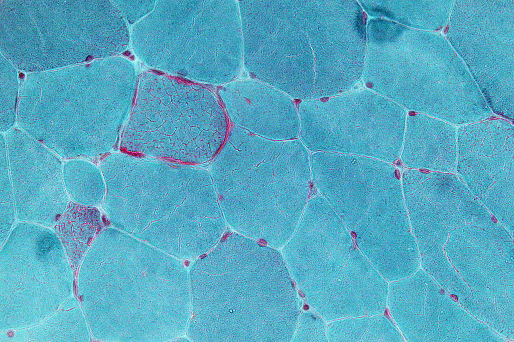
Jasmin discovered that her extreme fatigue, muscle pain, vision problems, and vomiting were due to problems in her mitochondria, like the damaged mitochondria shown in red in Figure 4.14.1. Mitochondria are small, membrane-bound organelles found in eukaryotic cells that provide energy for the cells of the body. They do this by carrying out the final two steps of aerobic cellular respiration: the Krebs cycle and electron transport. This is the major way that the human body breaks down the sugar glucose from food into a form of energy cells can use, namely the molecule ATP.
Because mitochondria provide energy for cells, you can understand why Jasmin was experiencing extreme fatigue, particularly after running. Her damaged mitochondria could not keep up with her need for energy, particularly after intense exercise, which requires a lot of additional energy. What is perhaps not so obvious are the reasons for her other symptoms, such as blurry vision, muscle spasms, and vomiting. All of the cells in the body require energy in order to function properly. Mitochondrial diseases can cause problems in mitochondria in any cell of the body, including muscle cells and cells of the nervous system, which includes the brain and nerves. The nervous system and muscles work together to control vision and digestive system functions, such as vomiting, so when they are not functioning properly, a variety of symptoms can emerge. This also explains why Jasmin’s niece, who has a similar mitochondrial disease, has symptoms related to brain function, such as seizures and learning disabilities. Our cells are microscopic, and mitochondria are even tinier — but they are essential for the proper functioning of our bodies. When they are damaged, serious health effects can occur.

One seemingly confusing aspect of mitochondrial diseases is that the type of symptoms, severity of symptoms, and age of onset can vary wildly between people — even within the same family! In Jasmin’s case, she did not notice symptoms until adulthood, while her niece had more severe symptoms starting at a much younger age. This makes sense when you know more about how mitochondrial diseases work.
Inherited mitochondrial diseases can be due to damage in either the DNA in the nucleus of cells or in the DNA in the mitochondria themselves. Recall that mitochondria are thought to have evolved from prokaryotic organisms that were once free-living, but were then infected or engulfed by larger cells. One of the pieces of evidence that supports this endosymbiotic theory is that mitochondria have their own, separate DNA. When the mitochondrial DNA is damaged (or mutated) it can result in some types of mitochondrial diseases. However, these mutations do not typically affect all of the mitochondria in a cell. During cell division, organelles such as mitochondria are replicated and passed down to the new daughter cells. If some of the mitochondria are damaged, and others are not, the daughter cells can have different amounts of damaged mitochondria. This helps explain the wide range of symptoms in people with mitochondrial diseases — even ones in the same family — because different cells in their bodies are affected in varying degrees. Jasmin’s niece was affected strongly and her symptoms were noticed early, while Jasmin’s symptoms were more mild and did not become apparent until adulthood.
There is still much more that needs to be discovered about the different types of mitochondrial diseases. But by learning about cells, their organelles, how they obtain energy, and how they divide, you should now have a better understanding of the biology behind these diseases.
Apply your understanding of cells to your own life. Can you think of other diseases that affect cellular structures or functions. Do they affect people you know? Since your entire body is made of cells, when cells are damaged or not functioning properly, it can cause a wide variety of health problems.
Chapter 4 Summary
Type your learning objectives here.
In this chapter you learned many facts about cells. Specifically, you learned that:
- Cells are the basic units of structure and function of living things.
- The first cells were observed from cork by Hooke in the 1600s. Soon after, van Leeuwenhoek observed other living cells.
- In the early 1800s, Schwann and Schleiden theorized that cells are the basic building blocks of all living things. Around 1850, Virchow saw cells dividing, and added his own theory that living cells arise only from other living cells. These ideas led to cell theory, which states that all organisms are made of cells, all life functions occur in cells, and all cells come from other cells.
- The invention of the electron microscope in the 1950s allowed scientists to see organelles and other structures inside cells for the first time.
- There is variation in cells, but all cells have a plasma membrane, cytoplasm, ribosomes, and DNA.
-
- The plasma membrane is composed mainly of a bilayer of phospholipid molecules and forms a barrier between the cytoplasm inside the cell and the environment outside the cell. It allows only certain substances to pass in or out of the cell. Some cells have extensions of their plasma membrane with other functions, such as flagella or cilia.
- Cytoplasm is a thick solution that fills a cell and is enclosed by the plasma membrane. It helps give the cell shape, holds organelles, and provides a site for many of the biochemical reactions inside the cell. The liquid part of the cytoplasm is called cytosol.
- Ribosomes are small structures where proteins are made.
- Cells are usually very small, so they have a large enough surface area-to-volume ratio to maintain normal cell processes. Cells with different functions often have different shapes.
- Prokaryotic cells do not have a nucleus. Eukaryotic cells have a nucleus, as well as other organelles. An organelle is a structure within the cytoplasm of a cell that is enclosed within a membrane and performs a specific job.
- The cytoskeleton is a highly organized framework of protein filaments and tubules that criss-cross the cytoplasm of a cell. It gives the cell shape and helps to hold cell structures (such as organelles) in place.
- The nucleus is the largest organelle in a eukaryotic cell. It is considered to be the cell’s control center, and it contains DNA and controls gene expression, including which proteins the cell makes.
- The mitochondrion is an organelle that makes energy available to cells. According to the widely accepted endosymbiotic theory, mitochondria evolved from prokaryotic cells that were once free-living organisms that infected or were engulfed by larger prokaryotic cells.
- The endoplasmic reticulum (ER) is an organelle that helps make and transport proteins and lipids. Rough endoplasmic reticulum (RER) is studded with ribosomes. Smooth endoplasmic reticulum (SER) has no ribosomes.
- The Golgi apparatus is a large organelle that processes proteins and prepares them for use both inside and outside the cell. It is also involved in the transport of lipids around the cell.
- Vesicles and vacuoles are sac-like organelles that may be used to store and transport materials in the cell or as chambers for biochemical reactions. Lysosomes and peroxisomes are vesicles that break down foreign matter, dead cells, or poisons.
- Centrioles are organelles located near the nucleus that help organize the chromosomes before cell division so each daughter cell receives the correct number of chromosomes.
- There are two basic ways that substances can cross the cell’s plasma membrane: passive transport (which requires no energy expenditure by the cell) and active transport (which requires energy).
- No energy is needed from the cell for passive transport because it occurs when substances move naturally from an area of higher concentration to an area of lower concentration. Types of passive transport in cells include:
-
- Simple diffusion, which is the movement of a substance due to differences in concentration without any help from other molecules. This is how very small, hydrophobic molecules, such as oxygen and carbon dioxide, enter and leave the cell.
- Osmosis, which is the diffusion of water molecules across the membrane.
- Facilitated diffusion, which is the movement of a substance across a membrane due to differences in concentration, but only with the help of transport proteins in the membrane (such as channel proteins or carrier proteins). This is how large or hydrophilic molecules and charged ions enter and leave the cell.
- Active transport requires energy to move substances across the plasma membrane, often because the substances are moving from an area of lower concentration to an area of higher concentration or because of their large size. Two examples of active transport are the sodium-potassium pump and vesicle transport.
-
- The sodium-potassium pump moves sodium ions out of the cell and potassium ions into the cell, both against a concentration gradient, in order to maintain the proper concentrations of both ions inside and outside the cell and to thereby control membrane potential.
- Vesicle transport uses vesicles to move large molecules into or out of cells.
- Energy is the ability to do work. It is needed by every living cell to carry out life processes.
- The form of energy that living things need is chemical energy, and it comes from food. Food consists of organic molecules that store energy in their chemical bonds.
- Autotrophs (producers) make their own food. Think of plants that make food by photosynthesis. Heterotrophs (consumers) obtain food by eating other organisms.
- Organisms mainly use the molecules glucose and ATP for energy. Glucose is the compact, stable form of energy that is carried in the blood and taken up by cells. ATP contains less energy and is used to power cell processes.
- The flow of energy through living things begins with photosynthesis, which creates glucose. The cells of organisms break down glucose and make ATP.
- Cellular respiration is the aerobic process by which living cells break down glucose molecules, release energy, and form molecules of ATP. Overall, this three-stage process involves glucose and oxygen reacting to form carbon dioxide and water.
-
- Glycolysis, the first stage of cellular respiration, takes place in the cytoplasm. In this step, enzymes split a molecule of glucose into two molecules of pyruvate, which releases energy that is transferred to ATP.
- Transition Reaction takes place between glycolysis and Krebs Cycle. It is a very short reaction in which the pyruvate molecules from glycolysis are converted into Acetyl CoA in order to enter the Krebs Cycle.
- Krebs Cycle, the second stage of cellular respiration, takes place in the matrix of a mitochondrion. During this stage, two turns through the cycle result in all of the carbon atoms from the two pyruvate molecules forming carbon dioxide and the energy from their chemical bonds being stored in a total of 16 energy-carrying molecules (including four from glycolysis).
- The Electron Transport System, he third stage of cellular respiration, takes place on the inner membrane of the mitochondrion. Electrons are transported from molecule to molecule down an electron-transport chain. Some of the energy from the electrons is used to pump hydrogen ions across the membrane, creating an electrochemical gradient that drives the synthesis of many more molecules of ATP.
- In all three stages of aerobic cellular respiration combined, as many as 38 molecules of ATP are produced from just one molecule of glucose.
- Some organisms can produce ATP from glucose by anaerobic respiration, which does not require oxygen. Fermentation is an important type of anaerobic process. There are two types: alcoholic fermentation and lactic acid fermentation. Both start with glycolysis.
-
- Alcoholic fermentation is carried out by single-celled organisms, including yeasts and some bacteria. We use alcoholic fermentation in these organisms to make biofuels, bread, and wine.
- Lactic acid fermentation is undertaken by certain bacteria, including the bacteria in yogurt, and also by our muscle cells when they are worked hard and fast.
- Anaerobic respiration produces far less ATP (typically produces 2 ATP) than does aerobic cellular respiration, but it has the advantage of being much faster.
- The cell cycle is a repeating series of events that includes growth, DNA synthesis, and cell division.
- In a eukaryotic cell, the cell cycle has two major phases: interphase and mitotic phase. During interphase, the cell grows, performs routine life processes, and prepares to divide. During mitotic phase, first the nucleus divides (mitosis) and then the cytoplasm divides (cytokinesis), which produces two daughter cells.
-
- Until a eukaryotic cell divides, its nuclear DNA exists as a grainy material called chromatin. After DNA replicates and the cell is about to divide, the DNA condenses and coils into the X-shaped form of a chromosome. Each chromosome consists of two sister chromatids, which are joined together at a centromere.
- During mitosis, sister chromatids separate from each other and move to opposite poles of the cell. This happens in four phases: prophase, metaphase, anaphase, and telophase.
- The cell cycle is controlled mainly by regulatory proteins that signal the cell to either start or delay the next phase of the cycle at key checkpoints.
- Cancer is a disease that occurs when the cell cycle is no longer regulated, often because the cell’s DNA has become damaged. Cancerous cells grow out of control and may form a mass of abnormal cells called a tumor.
In this chapter, you learned about cells and some of their functions, as well as how they pass genetic material in the form of DNA to their daughter cells. In the next chapter, you will learn how DNA is passed down to offspring, which causes traits to be inherited. These traits may be innocuous (such as eye colour) or detrimental (such as mutations that cause disease). The study of how genes are passed down to offspring is called genetics, and as you will learn in the next chapter, this is an interesting topic that is highly relevant to human health.
Chapter 4 Review
- Sequence:
- Drag and Drop:
- True or False:
- Multiple Choice:
- Briefly explain how the energy in the food you eat gets there, and how it provides energy for your neurons in the form necessary to power this process.
- Explain why the inside of the plasma membrane — the side that faces the cytoplasm of the cell — must be hydrophilic.
- Explain the relationships between interphase, mitosis, and cytokinesis.
Attributions
Figure 4.14.1
Mitochondrial Disease muscle sample by Nephron is used under a CC BY-SA 3.0 (https://creativecommons.org/licenses/by-sa/3.0) license.
Figure 4.14.2
Aunt and Niece by Tatiana Rodriguez on Unsplash is used under the Unsplash License (https://unsplash.com/license).
Reference
Wikipedia contributors. (2020, June 6). Mitochondrial disease. In Wikipedia. https://en.wikipedia.org/w/index.php?title=Mitochondrial_disease&oldid=961126371
A double-membrane-bound organelle found in most eukaryotic organisms. Mitochondria convert oxygen and nutrients into adenosine triphosphate (ATP). ATP is the chemical energy "currency" of the cell that powers the cell's metabolic activities.
Cells which have a nucleus enclosed within membranes, unlike prokaryotes, which have no membrane-bound organelles.
A series of chemical reactions used by all aerobic organisms to release stored energy through the oxidation of acetyl-CoA derived from carbohydrates, fats, and proteins.
A series of electron transporters embedded in the inner mitochondrial membrane that shuttles electrons from NADH and FADH2 to molecular oxygen. In the process, protons are pumped from the mitochondrial matrix to the intermembrane space, and oxygen is reduced to form water.
A complex organic chemical that provides energy to drive many processes in living cells, e.g. muscle contraction, nerve impulse propagation, and chemical synthesis. Found in all forms of life, ATP is often referred to as the "molecular unit of currency" of intracellular energy transfer.
Deoxyribonucleic acid - the molecule carrying genetic instructions for the development, functioning, growth and reproduction of all known organisms and many viruses.
An evolutionary theory of the origin of eukaryotic cells from prokaryotic organisms.
A semi-permeable lipid bilayer that separates the interior of all cells from their surroundings.
The jellylike material that makes up much of a cell inside the cell membrane, and, in eukaryotic cells, surrounds the nucleus. The organelles of eukaryotic cells, such as mitochondria, the endoplasmic reticulum, and (in green plants) chloroplasts, are contained in the cytoplasm.
A large complex of RNA and protein which acts as the site of RNA translation, building proteins from amino acids using messenger RNA as a template.
A whip-like structure that allows a cell to move.
Tiny hairlike organelles, identical in structure to flagella, that line the surfaces of certain cells and beat in rhythmic waves, providing locomotion to ciliate protozoans and moving liquids along internal epithelial tissue in animals.
Cells which lack membrane-bound structures, specifically a nucleus. Instead they generally have a single circular chromosome located in an area of the cell called the nucleoid.
A central organelle containing hereditary material.
A complex network of interlinking protein filaments that extends from the cell nucleus to the cell membrane, gives the cell its shape and help organize the cell's parts.
An organelle found in eukaryotic cells. Its main function is to produce proteins. It is a portion of the endoplasmic reticulum which is studded with attached ribosomes.
An organelle found in eukaryotic cells with the function of making cellular products such as hormones and lipids. The smooth endoplasmic reticulum is a part of the endoplasmic reticulum that does not have attached ribosomes.
A membrane-bound organelle found in eukaryotic cells made up of a series of flattened stacked pouches with the purpose of collecting and dispatching protein and lipid products received from the endoplasmic reticulum (ER). Also referred to as the Golgi complex or the Golgi body.
A structure within a cell, consisting of lipid bilayer. Vesicles form naturally during the processes of secretion, uptake and transport of materials within the plasma membrane.
A membrane-bound organelle which is present in all plant and fungal cells and some protist, animal and bacterial cells. It's function is storage of substances and to maintain the rigidity of plant cells.
A cylindrical organelle composed of microtubules located near the nucleus in animal cells, occurring in pairs and involved in the development of spindle fibers in cell division.
a type of movement of substances across the cell membrane which does not require energy because the substances are moving with the concentration gradient (from high to low concentration).
The movement of ions or molecules across a cell membrane into a region of higher concentration, assisted by enzymes and requiring energy.
The movement of a substance from an area of high concentration to an area of low concentration.
The movement of water or other solvent through a plasma membrane from a region of low solute concentration to a region of high solute concentration.
The passive movement of molecules across the cell membrane with the aid of a membrane protein.
An organism that produces complex organic compounds (such as carbohydrates, fats, and proteins) from simple substances present in its surroundings, generally using energy from light (photosynthesis) or inorganic chemical reactions (chemosynthesis).
An organism that cannot produce its own food, relying instead on the intake of nutrition from other sources of organic carbon, mainly plant or animal matter. In the food chain, heterotrophs are primary, secondary and tertiary consumers, but not producers.
Glucose (also called dextrose) is a simple sugar with the molecular formula C6H12O6. Glucose is the most abundant monosaccharide, a subcategory of carbohydrates. Glucose is mainly made by plants and most algae during photosynthesis from water and carbon dioxide, using energy from sunlight.
Photosynthesis is a process used by plants and other organisms to convert light energy into chemical energy that can later be released to fuel the organisms' activities.
A set of metabolic reactions and processes that take place in the cells of organisms to convert biochemical energy from nutrients into adenosine triphosphate (ATP).
The metabolic pathway that converts glucose C₆H₁₂O₆, into pyruvate. The free energy released in this process is used to form the high-energy molecules ATP and NADH. Glycolysis is a sequence of ten enzyme-catalyzed reactions.
Respiration using electron acceptors other than molecular oxygen. Although oxygen is not the final electron acceptor, the process still uses a respiratory electron transport chain.
A metabolic process that produces chemical changes in organic substrates through the action of enzymes. In biochemistry, it is narrowly defined as the extraction of energy from carbohydrates in the absence of oxygen.
A biological process which converts sugars such as glucose, fructose, and sucrose into cellular energy, producing ethanol and carbon dioxide as by-products.
A cycle of growth and division that cells go through. It includes interphase (G1, S, and G2) and the mitotic phase.
A group of diseases involving abnormal cell growth with the potential to invade or spread to other parts of the body.
