5.4 DNA Replication
By Christine Miller
DNA Replication: Overview
DNA replication is required for the growth or replication of an organism. You started as one single cell and are now made up of approximately 37 trillion cells! Each and every one of these cells contains the exact same copy of DNA, which originated from the first cell that was you. How did you get from one set of DNA, to 37 million sets, one for each cell? Through DNA replication.
Knowledge of DNA’s structure helped scientists understand DNA replication, the process by which DNA is copied. It occurs during the synthesis (S) phase of the eukaryotic cell cycle. DNA must be copied so that each new daughter cell will have a complete set of chromosomes after cell division occurs.
DNA replication is referred to as “semi-conservative”. What this means is when a strand of DNA is replicated, each of the two original strands acts as a template for a new complementary strand. When the replication process is complete, there are two identical sets of DNA, each containing one of the original strands of DNA, and one newly synthesized strand.
DNA replication involves a certain sequence of events. For each event, there is a specific enzyme which facilitates the process. There are four main enzymes that facilitate DNA replication: helicase, primase, DNA polymerase, and ligase.
DNA Replication: The Process
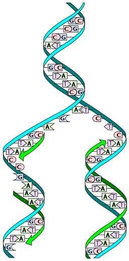
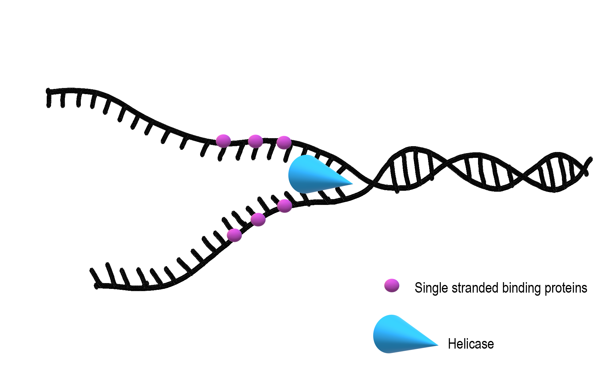
DNA replication begins when an enzyme called helicase unwinds, and unzips the DNA molecule. If you recall the structure of DNA, you may remember that it consists of two long strands of nucleotides held together by hydrogen bonds between complementary nitrogenous bases. This forms a ladder-like structure which is in a coiled shape. In order to start DNA replication, helicase needs to unwind the molecule and break apart the hydrogen bonds holding together complementary nitrogenous bases. This causes the two strands of DNA to separate.
Small molecules called single-stranded binding proteins (SSB) attach to the loose strands of DNA to keep them from re-forming the hydrogen bonds that helicase just broke apart.
Once the nitrogenous bases from the inside of the DNA molecule are exposed, the creation of a new, complementary strand can begin. DNA polymerase creates the new strand, but it needs some help in finding the correct place to begin, so primase lays down a short section of RNA primer (shown in green in Figure 5.4.3). Once this short section of primer is laid, DNA polymerase can bind to the DNA molecule and start connecting nucleotides in the correct order to match the sequence of nitrogenous bases on the template (original) strand.

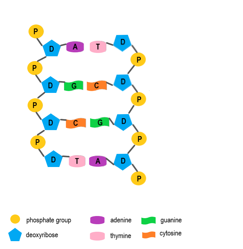
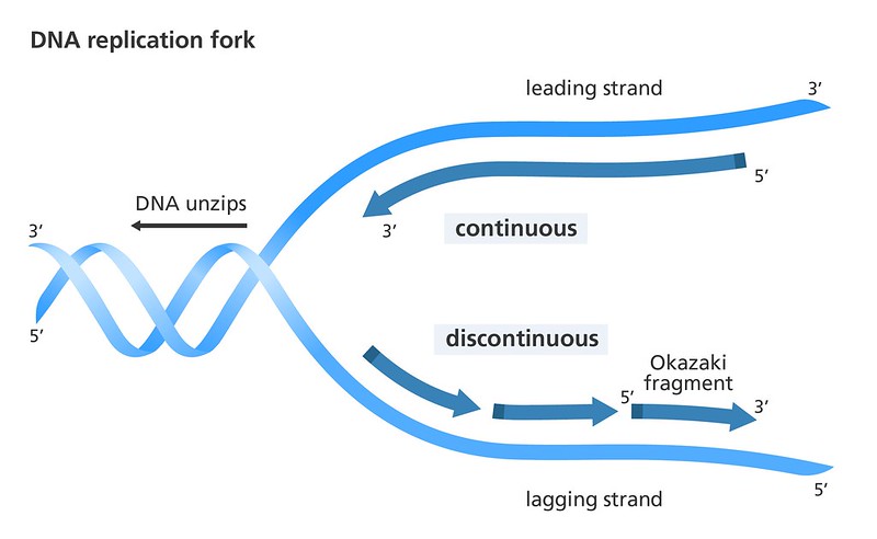
If we think about the DNA molecule, we may remember that the two strands of DNA run antiparallel to one another. This means that in the sugar-phosphate backbone, one strand of the DNA has the sugar oriented in the “up” position, and the other strand has the phosphate oriented in the “up” position (see Figure 5.4.4). DNA polymerase is an enzyme which can only work in one direction on the DNA molecule. This means that one strand of DNA can be replicated in one long string, as DNA polymerase follows helicase as it unzips the DNA molecule. This strand is termed the “leading strand”. The other strand, however, can only be replicated in small chunks since the DNA polymerase replicates in the opposite direction that helicase is unzipping. This strand is termed the “lagging strand”. These small chunks of replicated DNA on the lagging strand are called Okazaki fragments.
Take a look at Figure 5.4.5 and find the Okazaki fragments, the leading strand and the lagging strand.
Once DNA polymerase has replicated the DNA, a third enzyme called ligase completes the final stage of DNA replication, which is repairing the sugar-phosphate backbone. This connects the gaps in the backbone between Okazaki fragments. Once this is complete, the DNA coils back into its classic double helix structure.
Semi-Conservative Replication
When DNA replication is complete, there are two identical sets of double stranded DNA, each with one strand from the original, template, DNA molecule, and one strand that was newly synthesized during the DNA replication process. Because each new set of DNA contains one old and one new strand, we describe DNA as being semi-conservative.
Watch this video for a great overview of DNA replication:
DNA Replication (Updated), Amoeba Sisters, 2019.
5.4 Summary
- DNA replication requires the action of three main enzymes each with their own specific role:
- Helicase unzips and unwinds the DNA molecule.
- DNA polymerase creates a new complementary strand of DNA on each of the originals halves that were separated by helicase. New nucleotides are added through complementary base pairing: A pairs with T, and C with G.
- Ligase repairs gaps in the sugar-phosphate backbone between Okazaki fragments.
- DNA replication is semi-conservative because each daughter molecule contains one strand from the parent molecule and one new complementary strand.
5.4 Review Questions
2. Why are Okazaki fragments formed?
- Because helicase only unzips DNA in one direction.
- Because DNA is in a double helix.
- Because DNA polymerase only replicates DNA in one direction.
- Because DNA replication is semi-conservative.
3. Drag and drop to label the diagram.
5.4 Explore More
DNA replication – 3D, yourgenome, 2015.
Attributions
Figure 5.4.1
DNA_replication_split.svg by Madprime on Wikimedia Commons is used under a CC0 1.0
Public Domain Dedication license (https://creativecommons.org/publicdomain/zero/1.0/deed.en).
Figure 5.4.2
Helicase and single stranded binding proteins (1) by Christine Miller is used under a CC BY 4.0 (https://creativecommons.org/licenses/by/4.0/) license.
Figure 5.4.3
DNA polymerase and primase by Christine Miller is used under a CC BY 4.0 (https://creativecommons.org/licenses/by/4.0/) license.
Figure 5.4.4
DNA strands run antiparallel by Christine Miller is used under a CC BY 4.0 (https://creativecommons.org/licenses/by/4.0/) license.
Figure 5.4.5
Leading and lagging strand/ DNA Replication/ by yourgenome on Flickr is used under a CC BY-NC-SA 2.0 (https://creativecommons.org/licenses/by-nc-sa/2.0/) license.
References
Amoeba Sisters. (2019, June 28). DNA replication (Updated). YouTube. https://www.youtube.com/watch?v=Qqe4thU-os8&feature=youtu.be
Betts, J. G., Young, K.A., Wise, J.A., Johnson, E., Poe, B., Kruse, D.H., Korol, O., Johnson, J.E., Womble, M., DeSaix, P. (2013, April 25). Figure 3.24 DNA Replication [digital image]. In Anatomy and Physiology. OpenStax. https://openstax.org/books/anatomy-and-physiology/pages/3-3-the-nucleus-and-dna-replication CC BY 4.0 (https://creativecommons.org/licenses/by/4.0/)
yourgenome. (2015, June 26). DNA replication – 3D. YouTube. https://www.youtube.com/watch?v=TNKWgcFPHqw&feature=youtu.be
The process by which DNA is copied.
A cycle of growth and division that cells go through. It includes interphase (G1, S, and G2) and the mitotic phase.
The process by which a parent cell divides into two or more daughter cells. Cell division usually occurs as part of a larger cell cycle.
Biological molecules that lower amount the energy required for a reaction to occur.
