18.2 Introduction to the Reproductive System
Created by CK-12 Foundation/Adapted by Christine Miller
It’s All about Sex
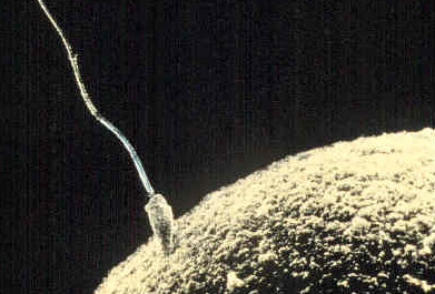
A tiny sperm from dad breaks through the surface of a huge egg from mom. Voilà! In nine months, a new son or daughter will be born. Like most other multicellular organisms, human beings reproduce sexually. In human sexual reproduction, males produce sperm and females produce eggs, and a new offspring forms when a sperm unites with an egg. How do sperm and eggs form? And how do they arrive together at the right place and time so they can unite to form a new offspring? These are functions of the reproductive system.
What Is the Reproductive System?
The reproductive system is the human organ system responsible for the production and fertilization of gametes (sperm or eggs) and, in females, the carrying of a fetus. Both male and female reproductive systems have organs called gonads that produce gametes. A gamete is a haploid cell that combines with another haploid gamete during fertilization, forming a single diploid cell called a zygote. Besides producing gametes, the gonads also produce sex hormones. Sex hormones are endocrine hormones that control the development of sex organs before birth, sexual maturation at puberty, and reproduction once sexual maturation has occurred. Other reproductive system organs have various functions, such as maturing gametes, delivering gametes to the site of fertilization, and providing an environment for the development and growth of an offspring.
Sex Differences in the Reproductive System
The reproductive system is the only human organ system that is significantly different between males and females. Embryonic structures that will develop into the reproductive system start out the same in males and females, but by birth, the reproductive systems have differentiated. How does this happen?
Sex Differentiation
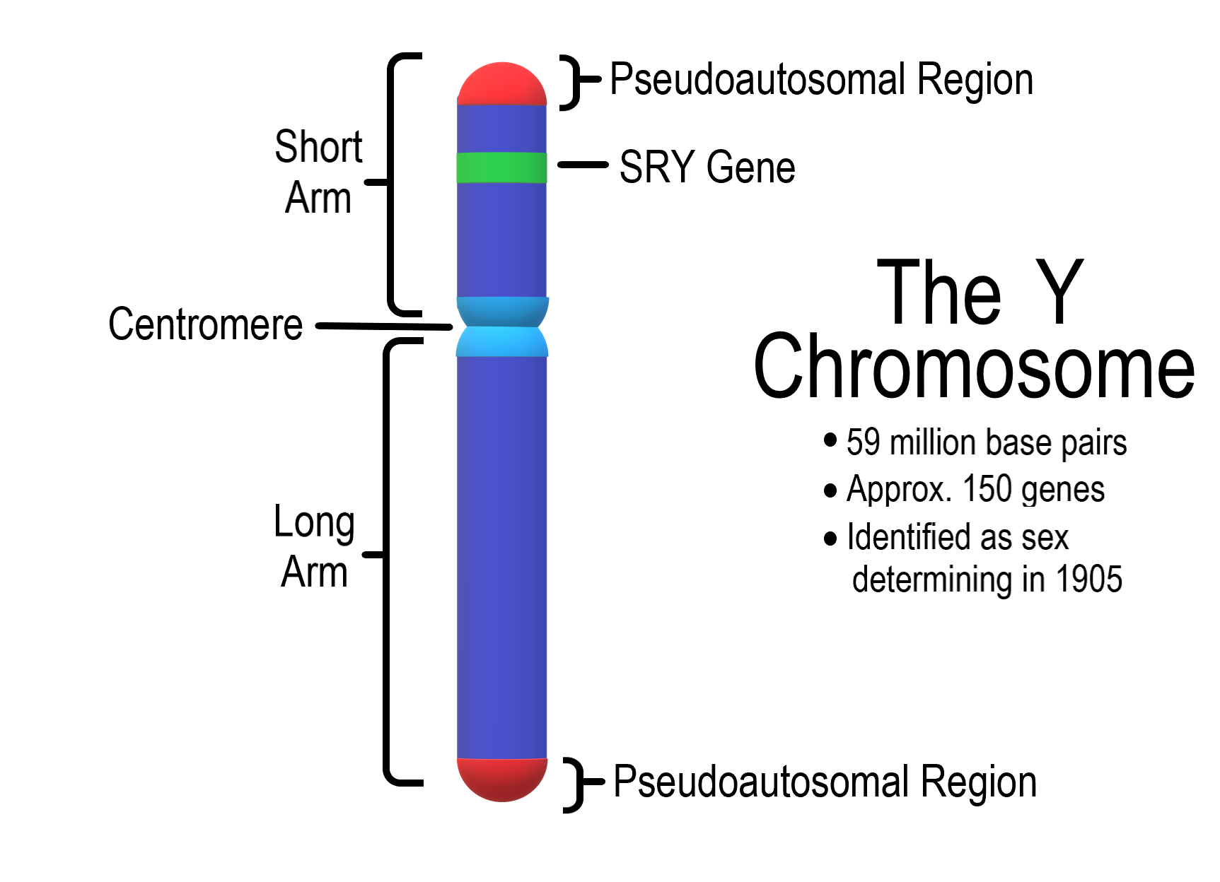
Starting around the seventh week after conception in genetically male (XY) embryos, a gene called SRY on the Y chromosome (shown in Figure 18.2.2) initiates the production of multiple proteins. These proteins cause undifferentiated gonadal tissue to develop into male gonads (testes). The male gonads then secrete hormones — including the male sex hormone testosterone — that trigger other changes in the developing offspring (now called a fetus), causing it to develop a complete male reproductive system. Without a Y chromosome, an embryo will develop female gonads (ovaries) that will produce the female sex hormone estrogen. Estrogen, in turn, will lead to the formation of the other organs of a normal female reproductive system.
Homologous Structures
Undifferentiated embryonic tissues develop into different structures in male and female fetuses. Structures that arise from the same tissues in males and females are called homologous structures. The male testes and female ovaries, for example, are homologous structures that develop from the undifferentiated gonads of the embryo. Likewise, the male penis and female clitoris are homologous structures that develop from the same embryonic tissues.
Sex Hormones and Maturation
Male and female reproductive systems are different at birth, but they are immature and incapable of producing gametes or sex hormones. Maturation of the reproductive system occurs during puberty, when hormones from the hypothalamus and pituitary gland stimulate the testes or ovaries to start producing sex hormones again. The main sex hormones are testosterone in males and estrogen in females. Sex hormones, in turn, lead to the growth and maturation of the reproductive organs, rapid body growth, and the development of secondary sex characteristics. Secondary sex characteristics are traits that are different in mature males and females, but are not directly involved in reproduction. They include facial hair in males and breasts in females.
Male Reproductive System
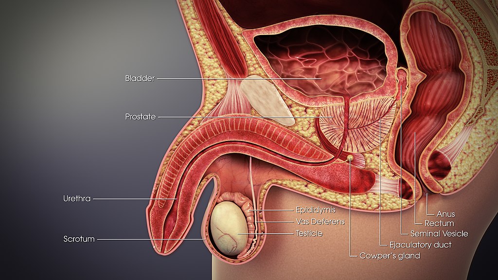
The main structures of the male reproductive system are external to the body and illustrated in Figure 18.2.3. The two testes (singular, testis) hang between the thighs in a sac of skin called the scrotum. The testes produce both sperm and testosterone. Resting atop each testis is a coiled structure called the epididymis (plural, epididymes). The function of the epididymes is to mature and store sperm. The penis is a tubular organ that contains the urethra and has the ability to stiffen during sexual arousal. Sperm passes out of the body through the urethra during a sexual climax (orgasm). This release of sperm is called ejaculation.
In addition to these organs, the male reproductive system consists of several ducts and glands that are internal to the body. The ducts, which include the vas deferens (also called the ductus deferens), transport sperm from the epididymis to the urethra. The glands, which include the prostate gland and seminal vesicles, produce fluids that become part of semen. Semen is the fluid that carries sperm through the urethra and out of the body. It contains substances that control pH and provide sperm with nutrients for energy.
Female Reproductive System
The main structures of the female reproductive system are internal to the body and shown in the following figure. They include the paired ovaries, which are small, ovoid structures that produce ova and secrete estrogen. The two oviducts (sometimes called Fallopian tubes or uterine tubes) start near the ovaries and end at the uterus. Their function is to transport ova from the ovaries to the uterus. If an egg is fertilized, it usually occurs while it is traveling through an oviduct. The uterus is a pear-shaped muscular organ that functions to carry a fetus until birth. It can expand greatly to accommodate a growing fetus, and its muscular walls can contract forcefully during labour to push the baby out of the uterus and into the vagina. The vagina is a tubular tract connecting the uterus to the outside of the body. The vagina is where sperm are usually deposited during sexual intercourse and ejaculation. The vagina is also called the birth canal because a baby travels through the vagina to leave the body during birth.
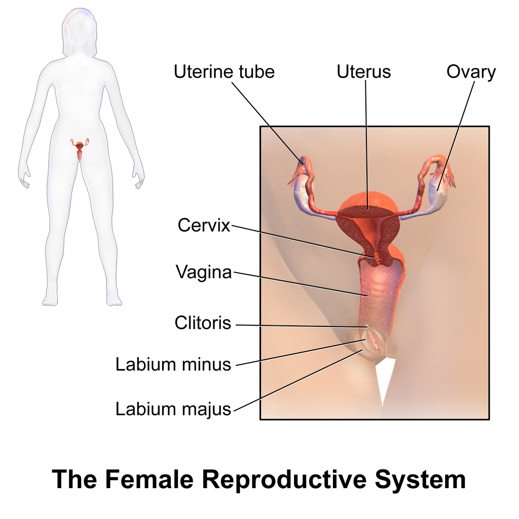
The external structures of the female reproductive system are referred to collectively as the vulva. They include the clitoris, which is homologous to the male penis. They also include two pairs of labia (singular, labium), which surround and protect the openings of the urethra and vagina.
18.2 Summary
- The reproductive system is the human organ system responsible for the production and fertilization of gametes and, in females, the carrying of a fetus.
- Both male and female reproductive systems have organs called gonads (testes in males, ovaries in females) that produce gametes (sperm or ova) and sex hormones (such as testosterone in males and estrogen in females). Sex hormones are endocrine hormones that control the prenatal development of reproductive organs, sexual maturation at puberty, and reproduction after puberty.
- The reproductive system is the only organ system that is significantly different between males and females. A Y-chromosome gene called SRY is responsible for undifferentiated embryonic tissues developing into a male reproductive system. Without a Y chromosome, the undifferentiated embryonic tissues develop into a female reproductive system.
- Structures such as testes and ovaries that arise from the same undifferentiated embryonic tissues in males and females are called homologous structures.
- Male and female reproductive systems are different at birth, but at that point, they are immature and nonfunctioning. Maturation of the reproductive system occurs during puberty, when hormones from the hypothalamus and pituitary gland stimulate the gonads to produce sex hormones again. The sex hormones, in turn, cause the changes of puberty.
- Male reproductive system organs include the testes, epididymis, penis, vas deferens, prostate gland, and seminal vesicles.
- Female reproductive system organs include the ovaries, oviducts, uterus, vagina, clitoris, and labia.
18.2 Review Questions
- What is the reproductive system?
-
- Explain the difference between the vulva and the vagina.
18.2 Explore More
Sex Determination: More Complicated Than You Thought, TED-Ed, 2012.
The evolution of animal genitalia – Menno Schilthuizen, TED-Ed, 2017.
Hormones and Gender Transition, Reactions, 2015.
Attributions
Figure 18.2.1
Sperm-egg by Unknown author on Wikimedia Commons is in the public domain (https://en.wikipedia.org/wiki/public_domain).
Figure 18.2.2
Y Chromosome by Christinelmiller on Wikimedia Commons is used under a CC BY-SA 4.0 (https://creativecommons.org/licenses/by-sa/4.0) license.
Figure 18.2.3
3D_Medical_Animation_Vas_Deferens by https://www.scientificanimations.com on Wikimedia Commons is used under a CC BY-SA 4.0 (https://creativecommons.org/licenses/by-sa/4.0) license.
Figure 18.2.4
Blausen_0399_FemaleReproSystem_01 by BruceBlaus on Wikimedia Commons is used under a CC BY 3.0 (https://creativecommons.org/licenses/by/3.0) license.
References
Blausen.com Staff. (2014). Medical gallery of Blausen Medical 2014. WikiJournal of Medicine 1 (2). DOI:10.15347/wjm/2014.010. ISSN 2002-4436.
Reactions. (2015, June 8). Hormones and gender transition. YouTube. https://www.youtube.com/watch?v=l5knvmy1Z3s&feature=youtu.be
TED-Ed. (2012, April 23). Sex determination: More complicated than you thought. YouTube. https://www.youtube.com/watch?v=kMWxuF9YW38&feature=youtu.be
TED-Ed. (2017, April 24). The evolution of animal genitalia – Menno Schilthuizen. YouTube. https://www.youtube.com/watch?v=vcPJkz-D5II&feature=youtu.be
The male reproductive cell.
The body system by which humans reproduce and bear live offspring.
One of a pair of organs that secrete sex hormones and produce gametes; testis in males and ovary in females.
A mature haploid male or female germ cell which is able to unite with another of the opposite sex in sexual reproduction to form a zygote.
The term used when a cell has half the usual number of chromosomes.
The fusion of haploid gametes, egg and sperm, to form the diploid zygote.
The union of the sperm cell and the egg cell. Also known as a fertilized ovum, the zygote begins as a single cell but divides rapidly in the days following fertilization. After this two-week period of cell division, the zygote eventually becomes an embryo.
An endocrine hormone secreted mainly by gonads that controls sexual development and reproduction.
An unborn offspring of a mammal, in particular an unborn human baby more than eight weeks after conception.
Structures that are similar in related species because it was inherited from a common ancestor; or structure that develops from the same undifferentiated embryonic tissue in males and females of the same species, such as the testis and ovary in humans.
A part of the brain that secretes hormones and connects the brain with the endocrine system.
The master gland of the endocrine system that secretes many hormones, the majority of which regulate other endocrine glands.
The male sex hormone secreted mainly by the testes.
The female sex hormone secreted mainly by the ovaries.
A trait that is different in males and females but is not directly involved in reproduction, such as male facial hair and female breasts.
Two male reproductive organs that produce sperm and secrete testosterone; male gonad.
A pouch-like external structure of the male reproductive system, located behind the penis, that contains the testes, epididymes, and part of the vas deferens.
One of two male reproductive organs where sperm mature and are stored until they leave the body during ejaculation.
The male reproductive organ containing the urethra, through which semen and urine pass out of the body.
One of a pair of thin tubes that transports sperm from an epididymis to an ejaculatory duct during ejaculation; also called sperm duct.
A tube-like organ of the urinary system that carries urine out of the body from the bladder and, in males, also carries semen out of the body.
A gland in the male reproductive system that secretes fluid into semen and provides nourishing substances to sperm.
One of a pair of glands of the male reproductive system that secretes fluid into semen.
Fluid containing sperm and glandular secretions, which nourishes sperm and carries them through the urethra and out of the body.
A pair of female reproductive organs that produces eggs and secretes estrogen.
The gamete produced by a female.
One of two female reproductive organs that carry eggs from an ovary to the uterus and are the site where fertilization usually takes place.
The female reproductive organ in which first an embryo and then a fetus grows and develops until birth.
The female reproductive organ that receives sperm during sexual intercourse and provides a passageway for a baby to leave the mother’s body during birth.
The physical activity of sex between two people.
The process in males in which muscle contractions propel sperm from the epididymes and out through the urethra in semen.
External female reproductive structures, including the clitoris, labia, and vaginal and urethral openings.
The small, sensitive external female organ that is part of the vulva and may lead to sexual arousal and/or orgasm when stimulated.
The “lips” of the vulva, consisting of folds of tissue that protect the urethral and vaginal openings.
A hormone is a signaling molecule produced by glands in multicellular organisms that target distant organs to regulate physiology and behavior.
A period during which humans become sexually mature.
