4.4 Plasma Membrane
Created by: CK-12/Adapted by Christine Miller
A Bag Full of Jell-O
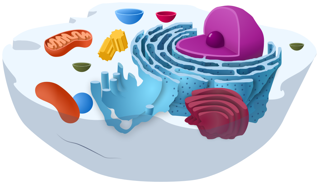
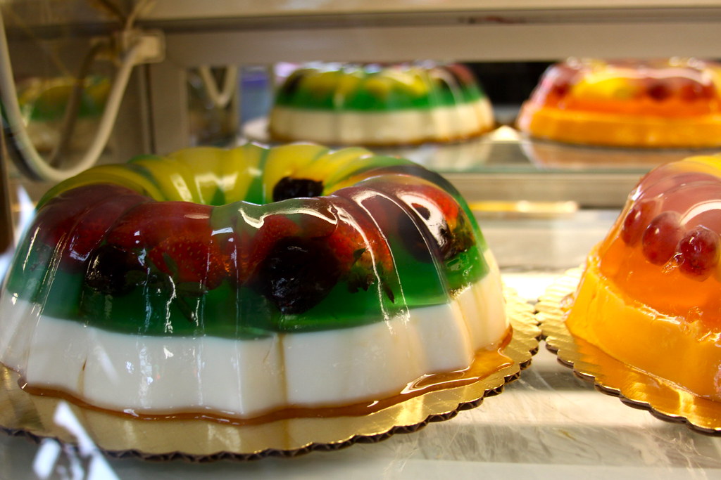
The simple cut-away model of an animal cell (Figure 4.4.1) shows that a cell resembles a plastic bag full of Jell-O. Its basic structure is a plasma membrane filled with cytoplasm. Like Jell-O containing mixed fruit (Figure 4.4.2), the cytoplasm of the cell also contains various structures, including a nucleus and other organelles. Your body is composed of trillions of cells, but all of them perform the same basic life functions. They all obtain and use energy, respond to the environment, and reproduce. How do your cells carry out these basic functions and keep themselves — and you — alive? To answer these questions, you need to know more about the structures that make up cells, starting with the plasma membrane.
What is the Plasma Membrane?
The plasma membrane is a structure that forms a barrier between the cytoplasm inside the cell and the environment outside the cell. Without the plasma membrane, there would be no cell. Although it is very thin and flexible, the plasma membrane protects and supports the cell by controlling everything that enters and leaves it. It allows only certain substances to pass through, while keeping others in or out. To understand how the plasma membrane controls what passes into or out of the cell, you need to know its basic structure.
Phospholipid Bilayer
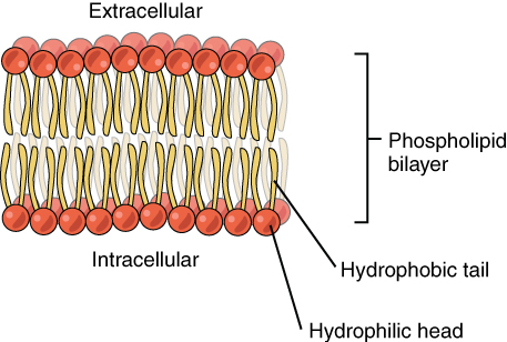
The plasma membrane is composed mainly of phospholipids, which consist of fatty acids and alcohol. The phospholipids in the plasma membrane are arranged in two layers, called a phospholipid bilayer. As shown in the simplified diagram in Figure 4.4.3, each individual phospholipid molecule has a phosphate group head (in red) and two fatty acid tails (in yellow). The head “loves” water (hydrophilic) and the tails “hate” water (hydrophobic). The water-hating tails are on the interior of the membrane, whereas the water-loving heads point outward, toward either the cytoplasm (intracellular) or the fluid that surrounds the cell (extracellular).
Hydrophobic molecules can easily pass through the plasma membrane if they are small enough, because they are water-hating like the interior of the membrane. Hydrophilic molecules, on the other hand, cannot pass through the plasma membrane — at least not without help — because they are water-loving like the exterior of the membrane.
Other Molecules in the Plasma Membrane
The plasma membrane also contains other molecules, primarily other lipids and proteins. The yellow molecules in the diagram here, for example, are the lipid cholesterol. Molecules of the steroid lipid cholesterol help the plasma membrane keep its shape. Proteins in the plasma membrane (shown blue in Figure 4.4.4) include: transport proteins that assist other substances in crossing the cell membrane, receptors that allow the cell to respond to chemical signals in its environment, and cell-identity markers that indicate what type of cell it is and whether it belongs in the body.

Additional Functions of the Plasma Membrane
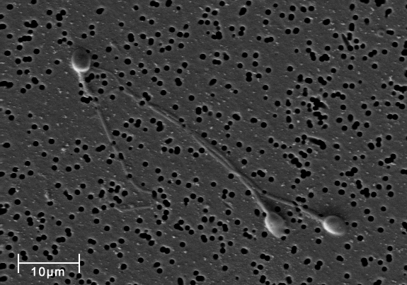
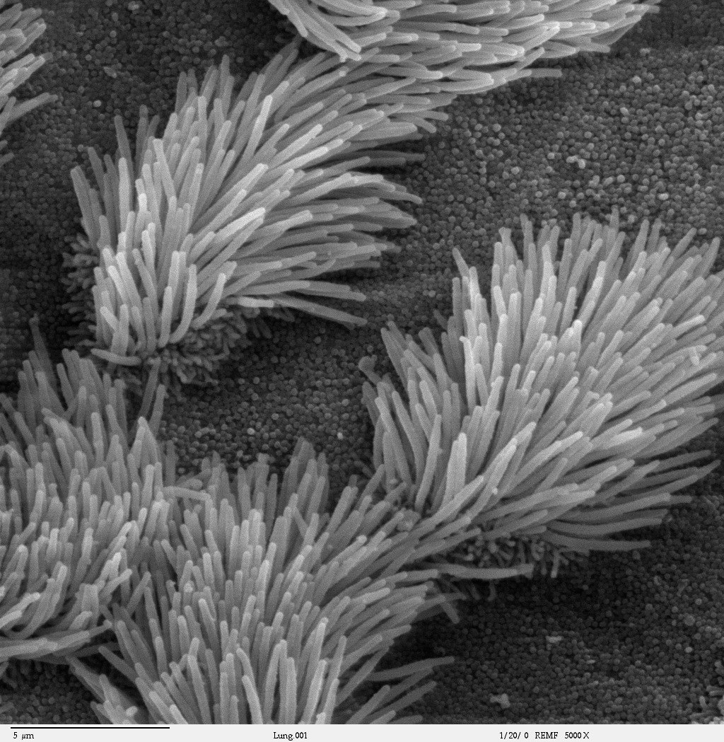
The plasma membrane may have extensions, such as whip-like flagella (singular flagellum) or brush-like cilia (singular cilium), shown below (Figure 4.4.5), that give it other functions. In single-celled organisms, these membrane extensions may help the organisms move. In multicellular organisms, the extensions have different functions. For example, the cilia on human lung cells sweep foreign particles and mucus toward the mouth and nose, while the flagellum on a human sperm cell allows it to swim.
Feature: My Human Body
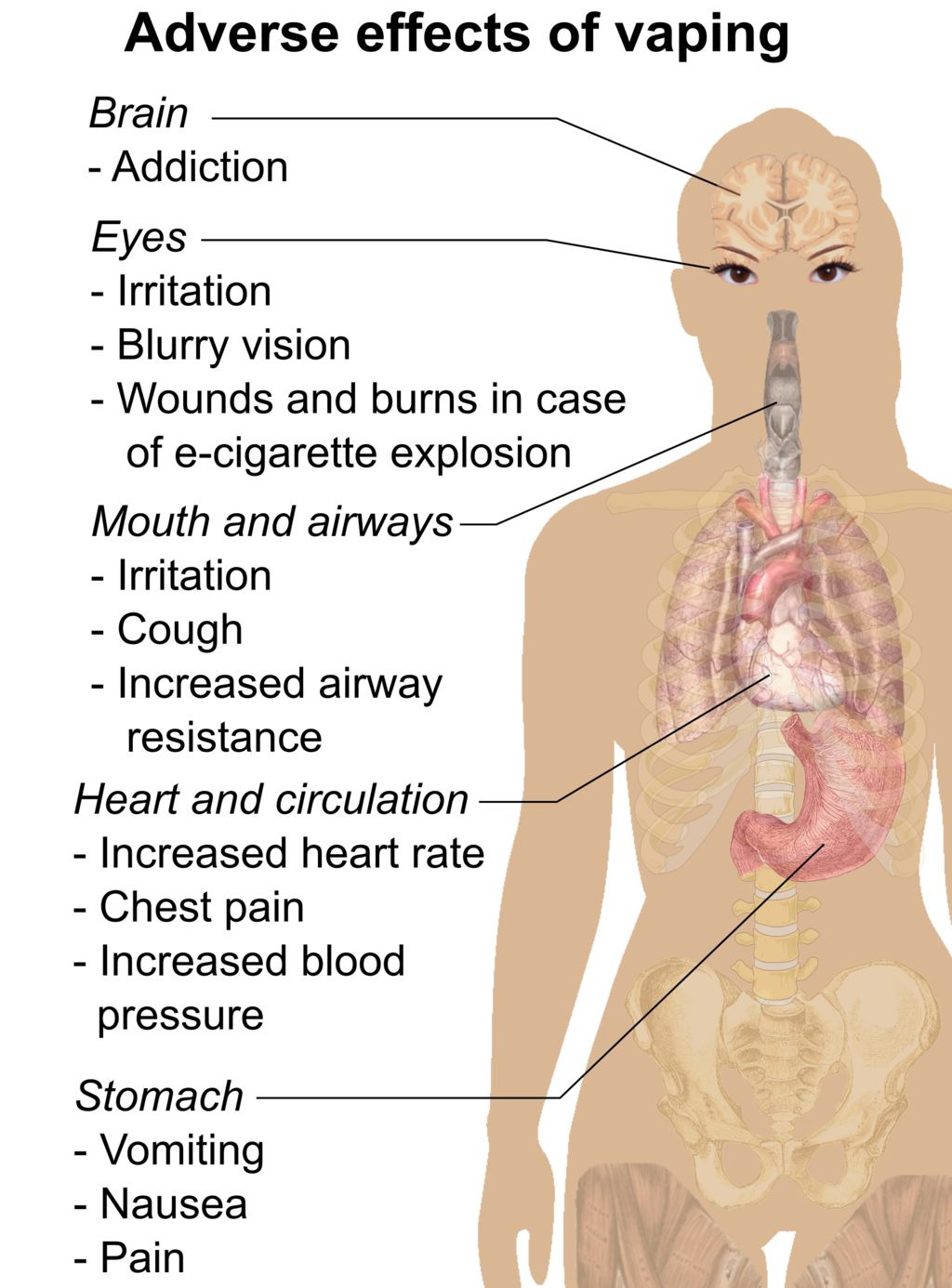
If you smoke or use e-cigarettes (vaping) and need another reason to quit, here’s a good one. We usually think of lung cancer as the major disease caused by smoking. But smoking and vaping can have devastating effects on the body’s ability to protect itself from repeated, serious respiratory infections, such as bronchitis and pneumonia.
Cilia are microscopic, hair-like projects on cells that line the respiratory, reproductive, and digestive systems. Cilia in the respiratory system line most of your airways, where they have the job of trapping and removing dust, germs, and other foreign particles before they can make you sick. Cilia secrete mucus that traps particles, and they move in a continuous wave-like motion that sweeps the mucus and particles upward toward the throat, where they can be expelled from the body. When you are sick and cough up phlegm, that’s what you are doing.
Smoking prevents cilia from performing these important functions. Chemicals in tobacco smoke paralyze the cilia so they can’t sweep mucus out of the airways. Those chemicals also inhibit the cilia from producing mucus. Fortunately, these effects start to wear off soon after the most recent exposure to tobacco smoke. If you stop smoking, your cilia will return to normal. Even if prolonged smoking has destroyed cilia, they will regrow and resume functioning in a matter of months after you stop smoking.
4.4 Summary
- The plasma membrane is a structure that forms a barrier between the cytoplasm inside the cell and the environment outside the cell. It allows only certain substances to pass in or out of the cell.
- The plasma membrane is composed mainly of a bilayer of phospholipid molecules. It also contains other molecules, such as the steroid cholesterol, which helps the membrane keep its shape, and transport proteins, which help substances pass through the membrane.
- The plasma membranes of some cells have extensions that have other functions, like flagella to help sperm move, or cilia to help keep our airways clear.
4.4 Review Questions
- What are the general functions of the plasma membrane?
- Describe the phospholipid bilayer of the plasma membrane.
- Identify other molecules in the plasma membrane. State their functions.
- Why do some cells have plasma membrane extensions, like flagella and cilia?
- Explain why hydrophilic molecules cannot easily pass through the cell membrane. What type of molecule in the cell membrane might help hydrophilic molecules pass through it?
- Which part of a phospholipid molecule in the plasma membrane is made of fatty acid chains? Is this part hydrophobic or hydrophilic?
- The two layers of phospholipids in the plasma membrane are called a phospholipid ____________.
-
- Steroid hormones can pass directly through cell membranes. Why do you think this is the case?
- Some antibiotics work by making holes in the plasma membrane of bacterial cells. How do you think this kills the cells?
- What is the name of the long, whip-like extensions of the plasma membrane that helps some single-celled organisms move?
4.4 Explore More
Insights into cell membranes via dish detergent – Ethan Perlstein, TED-Ed, 2013.
Inside the cell membrane, by The Amoeba Sisters, 2018.
Attributions
Figure 4.4.1
Animal Cell Unannotated, by Kelvin Song on Wikimedia Commons is used under a CC0 1.0 (https://creativecommons.org/publicdomain/zero/1.0/deed.en) public domain dedication license.
Figure 4.4.2
Jello mold at the mexican bakery photo by Aimée Knight on Flickr is used under a CC BY 2.0 (https://creativecommons.org/licenses/by/2.0/) license.
Figure 4.4.3
Phospholipid_Bilayer by OpenStax on Wikimedia Commons is used under a CC BY 4.0 (https://creativecommons.org/licenses/by/4.0) license.
Figure 4.4.4
Lipid bilayer by OpenStax on Wikimedia Commons is used under a CC BY 4.0 (https://creativecommons.org/licenses/by/4.0) license.
Figure 4.4.5
Spermatozoa-human-3140x by No specific author on Wikimedia Commons is released into the public domain (https://en.wikipedia.org/wiki/Public_domain).
Figure 4.4.6
Cilia/ Bronchiolar epithelium 3 – SEM by Charles Daghlian on Wikimedia Commons is released into the public domain (https://en.wikipedia.org/wiki/Public_domain).
Figure 4.4.7
Adverse effects of vaping (raster) by Mikael Häggström on Wikimedia Commons is released into the public domain (https://en.wikipedia.org/wiki/Public_domain).
References
Amoeba Sisters. (2018, February 27). Inside the cell membrane. YouTube. https://www.youtube.com/watch?v=qBCVVszQQNs&feature=youtu.be
Betts, J.G., Young, K.A., Wise, J.A., Johnson, E., Poe, B., Kruse, D.H., Korol, O., Johnson, J.E.. Womble, M., DeSaix. P. (2013, April 25). Figure 3.3 Phospolipid Bilayer [digital image]. In Anatomy and Physiology. OpenStax. https://openstax.org/books/anatomy-and-physiology/pages/3-1-the-cell-membrane
Betts, J.G., Young, K.A., Wise, J.A., Johnson, E., Poe, B., Kruse, D.H., Korol, O., Johnson, J.E.. Womble, M., DeSaix. P. (2013, April 25). Figure 3.4 Cell Membrane [digital image]. In Anatomy and Physiology. OpenStax. https://openstax.org/books/anatomy-and-physiology/pages/3-1-the-cell-membrane
Ghosh, A., Coakley, R. C., Mascenik, T., Rowell, T. R., Davis, E. S., Rogers, K., Webster, M. J., Dang, H., Herring, L. E., Sassano, M. F., Livraghi-Butrico, A., Van Buren, S. K., Graves, L. M., Herman, M. A., Randell, S. H., Alexis, N. E., & Tarran, R. (n.d.). Chronic E-Cigarette Exposure Alters the Human Bronchial Epithelial Proteome. American Journal of Respiratory and Critical /Care Medicine, 198(1), 67–76. https://doi-org.ezproxy.tru.ca/10.1164/rccm.201710-2033OC
TED-Ed. (2013, February 26). Insights into cell membranes via dish detergent – Ethan Perlstein. YouTube. https://www.youtube.com/watch?v=yAXnYcUjn5k&feature=youtu.be
The smallest unit of life, consisting of at least a membrane, cytoplasm, and genetic material.
A semi-permeable lipid bilayer that separates the interior of all cells from their surroundings.
The jellylike material that makes up much of a cell inside the cell membrane, and, in eukaryotic cells, surrounds the nucleus. The organelles of eukaryotic cells, such as mitochondria, the endoplasmic reticulum, and (in green plants) chloroplasts, are contained in the cytoplasm.
A central organelle containing hereditary material.
A tiny cellular structure that performs specific functions within a cell.
The ability to do work.
The production of offspring by sexual or asexual process.
A thin polar membrane made of two layers of phospholipid molecules. These membranes are flat sheets that form a continuous barrier around all cells.
Attracted to water.
Repelled by water.
A substance that is insoluble in water. Examples include fats, oils and cholesterol. Lipids are made from monomers such as glycerol and fatty acids.
A class of biological molecule consisting of linked monomers of amino acids and which are the most versatile macromolecules in living systems and serve crucial functions in essentially all biological processes.
A whip-like structure that allows a cell to move.
Tiny hairlike organelles, identical in structure to flagella, that line the surfaces of certain cells and beat in rhythmic waves, providing locomotion to ciliate protozoans and moving liquids along internal epithelial tissue in animals.
A group of diseases involving abnormal cell growth with the potential to invade or spread to other parts of the body.

.png)