7.9 Case Study Conclusion: Under Pressure
Created by CK-12 Foundation/Adapted by Christine Miller
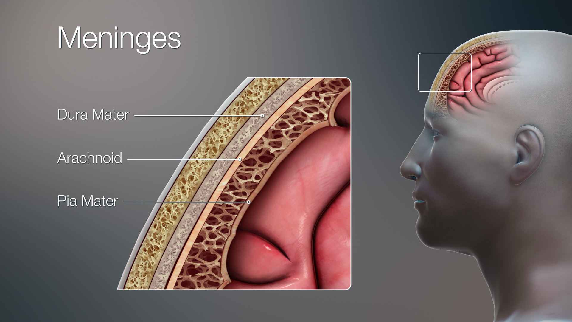
As you learned in this chapter, the human body consists of many complex systems that normally work together efficiently — like a well-oiled machine — to carry out life’s functions. For example, the image above (Figure 7.9.1) illustrates how the brain and spinal cord are protected by layers of membrane called meninges and fluid that flows between the meninges and in spaces called ventricles inside the brain. This fluid is called cerebrospinal fluid, and as you have learned, one of its important functions is to cushion and protect the brain and spinal cord, which make up most of the central nervous system (CNS). Additionally, cerebrospinal fluid circulates nutrients and removes waste products from the CNS. Cerebrospinal fluid is produced continually in the ventricles, circulates throughout the CNS, and is then reabsorbed by the bloodstream. If too much cerebrospinal fluid is produced, its flow is blocked, or not enough is reabsorbed, the system becomes out of balance and it can build up in the ventricles. This causes an enlargement of the ventricles called hydrocephalus that can put pressure on the brain, resulting in the types of neurological problems that former professional football player Jayson, described in the beginning of this chapter, is suffering from.
Recall that Jayson’s symptoms included loss of bladder control, memory loss, and difficulty walking. The cause of his symptoms was not immediately clear, although his doctors suspected that it related to the nervous system, since the nervous system acts as the control centre of the body, controlling and regulating many other organ systems. Jayson’s memory loss directly implicated the brain’s involvement, since that is the site of thoughts and memory. The urinary system is also controlled in part by the nervous system, so the inability to hold urine appropriately can also be a sign of a neurological issue. Jayson’s trouble walking involved the muscular system, which works alongside the skeletal system to enable movement of the limbs. In turn, the contraction of muscles is regulated by the nervous system. You can see why a problem in the nervous system can cause a variety of different symptoms by affecting multiple organ systems in the human body.
To try to find the exact cause of Jayson’s symptoms, his doctors performed a lumbar puncture (or spinal tap), which is the removal of some cerebrospinal fluid through a needle inserted into the lower part of the spinal canal. They then analyzed Jayson’s cerebrospinal fluid for the presence of pathogens (such as bacteria) to determine whether an infection was the cause of his neurological symptoms. When no evidence of infection was found, they used an MRI to observe the structures of his brain. This is when they discovered his enlarged ventricles, which are a hallmark of hydrocephalus.
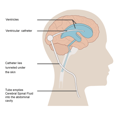
To treat Jayson’s hydrocephalus, a surgeon implanted a device called a shunt in his brain to remove the excess fluid. An illustration of a brain shunt is shown in Figure 9.7.2 . One side of the shunt consists of a small tube, called a catheter, which was inserted into Jayson’s ventricles. Excess cerebrospinal fluid is then drained through a one-way valve to the other end of the shunt, which was threaded under his skin to his abdominal cavity, where the fluid is released and can be reabsorbed by the bloodstream.
Implantation of a shunt is the most common way to treat hydrocephalus, and for some people, it can allow them to recover almost completely. However, there can be complications associated with a brain shunt. The shunt can have mechanical problems or cause an infection. Also, the rate of draining must be carefully monitored and adjusted to balance the rate of cerebrospinal fluid removal with the rate of its production. If it is drained too fast, it is called overdraining, and if it is drained too slowly, it is called underdraining. In the case of underdraining, the pressure on the brain and associated neurological symptoms will persist. In the case of overdraining, the ventricles can collapse, which can cause serious problems, such as the tearing of blood vessels and hemorrhaging. To avoid these problems, some shunts have an adjustable pressure valve, where the rate of draining can be adjusted by placing a special magnet over the scalp. You can see how the proper balance between cerebrospinal fluid production and removal is so critical – both in the causes of hydrocephalus and in its treatment.
In what other ways does your body regulate balance, or maintain a state of homeostasis? In this chapter you learned about the feedback loops that keep body temperature and blood glucose within normal ranges. Other important examples of homeostasis in the human body are the regulation of the pH in the blood and the balance of water in the body. You will learn more about homeostasis in different body systems in the coming chapters.
Thanks to Jayson’s shunt, his symptoms are starting to improve, but he has not fully recovered. Time may tell whether the removal of the excess cerebrospinal fluid from his ventricles will eventually allow him to recover normal functioning or whether permanent damage to his nervous system has already been done. The flow of cerebrospinal fluid might seem simple, but when it gets out of balance, it can easily wreak havoc on multiple organ systems because of the intricate interconnectedness of the systems within the human “machine.”
To learn more about hydrocephalus and its treatment, watch this video from Boston Children’s Hospital:
Hydrocephalus and its treatment | Boston Children’s Hospital, 2011.
Chapter 7 Summary
This chapter provided an overview of the organization and functioning of the human body. You learned that:
- The human body consists of multiple parts that function together to maintain life. The biology of the human body incorporates the body’s structure — or anatomy — and the body’s functioning, or physiology.
- The organization of the human body is a hierarchy of increasing size and complexity, starting at the level of atoms and molecules and ending at the level of the entire organism.
- Cells are the level of organization above atoms and molecules, and they are the basic units of structure and function of the human body. Each cell carries out basic life functions, as well as other specific roles. Cells of the human body show a lot of variation.
-
- Variations in cell function are generally reflected in variations in cell structure.
- Some cells are unattached to other cells and can move freely. Others are attached to each other and cannot move freely. Some cells can divide readily and form new cells, and others can divide only under exceptional circumstances. Many cells are specialized to produce and secrete particular substances.
- All the different cell types within an individual have the same genes. Cells can vary because different genes are expressed depending on the cell type.
- Many common types of human cells consist of several subtypes of cells, each of which has a special structure and function. For example, subtypes of bone cells include osteocytes, osteoblasts, osteogenic cells, and osteoclasts.
- A tissue is a group of connected cells that have a similar function. There are four basic types of human tissues that make up all the organs of the human body: epithelial, muscle, nervous, and connective tissues.
-
- Connective tissues, such as bone, tendons and blood, are made up of a scattering of living cells that are separated by non-living material, called extracellular matrix.
- Epithelial tissues, such as skin and mucous membranes, protect the body and its internal organs and secrete or absorb substances.
- Muscular tissues are made up of cells that have the unique ability to contract. They include skeletal, smooth, and cardiac muscle tissues.
- Nervous tissues are made up of neurons, which transmit messages, and neuroglia of various types, which play supporting roles.
- An organ is a structure that consists of two or more types of tissues that work together to do the same job. The brain and the heart are two examples.
-
- Many organs are composed of a major tissue that performs the organ’s main function, as well as other tissues that play supporting roles.
- The human body contains five organs that are considered vital for survival: the heart, brain, kidneys, liver, and lungs. If any of these five organs stops functioning, death of the organism is imminent without medical intervention.
- An organ system is a group of organs that work together to carry out a complex overall function. For example, the skeletal system provides structure to the body and protects internal organs.
-
- There are 11 major organ systems in the human organism. They are the integumentary, skeletal, muscular, nervous, endocrine, cardiovascular, lymphatic, respiratory, digestive, urinary, and reproductive systems. Only the reproductive system varies significantly between males and females.
- The human body is divided into a number of body cavities. A body cavity is a fluid-filled space in the body that holds and protects internal organs. The two largest human body cavities are the ventral cavity and dorsal cavity.
-
- The ventral cavity is at the anterior (or front) of the trunk. It is subdivided into the thoracic cavity, abdominal cavity and the pelvic cavity.
- The dorsal cavity is at the posterior (or back) of the body, and includes the head and the back of the trunk. It is subdivided into the cranial cavity and spinal cavity.
- Organ systems of the human body must work together to keep the body alive and functioning normally. This requires communication among organ systems. This is controlled by the autonomic nervous system and endocrine system. The autonomic nervous system controls involuntary body functions, such as heart rate and digestion. The endocrine system secretes hormones into the blood that travel to body cells and influence their activities.
-
- Cellular respiration is a good example of organ system interactions, because it is a basic life process that occurs in all living cells. It is the intracellular process that breaks down glucose with oxygen to produce carbon dioxide and energy. Cellular respiration requires the interaction of the digestive, cardiovascular, and respiratory systems.
- The fight-or-flight response is a good example of how the nervous and endocrine systems control other organ system responses. It is triggered by a message from the brain to the endocrine system and prepares the body for flight or a fight. Many organ systems are stimulated to respond, including the cardiovascular, respiratory, and digestive systems.
- Playing softball or doing other voluntary physical activities may involve the interaction of nervous, muscular, skeletal, respiratory, and cardiovascular systems.
- Homeostasis is the condition in which a system such as the human body is maintained in a more or less steady state. It is the job of cells, tissues, organs, and organ systems throughout the body to maintain homeostasis.
-
- For any given variable (such as body temperature), there is a particular set point that is the physiological optimum value. The spread of values around the set point that is considered insignificant is called the normal range.
- Homeostasis is generally maintained by a negative feedback loop that includes a stimulus, sensor, control centre, and effector. Negative feedback serves to reduce an excessive response and to keep a variable within the normal range. Negative feedback loops control body temperature and the blood glucose level.
- Sometimes homeostatic mechanisms fail, resulting in homeostatic imbalance. Diabetes is an example of a disease caused by homeostatic imbalance. Aging can bring about a reduction in the efficiency of the body’s control system, making the elderly more susceptible to disease.
- Positive feedback loops are not common in biological systems. Positive feedback serves to intensify a response until an end point is reached. Positive feedback loops control blood clotting and childbirth.
The severe and broad impact of hydrocephalus on the body’s systems highlights the importance of the nervous system and its role as the master control system of the body. In the next chapter, you will learn much more about the structures and functioning of this fascinating and important system.
Chapter 7 Review
-
- Compare and contrast tissues and organs.
-
-
- Which type of tissue lines the inner and outer surfaces of the body?
- What is a vital organ? What happens if a vital organ stops working?
- Name three organ systems that transport or remove wastes from the body.
- Name two types of tissue in the digestive system.
-
- Describe one way in which the integumentary and cardiovascular systems work together to regulate homeostasis in the human body.
-
- True or False: Body cavities are filled with air.
- In which organ system is the pituitary gland? Describe how the pituitary gland increases metabolism.
- When the level of thyroid hormone in the body gets too high, it acts on other cells to reduce production of more thyroid hormone. What type of feedback loop does this represent?
- Hypothetical organ A is the control centre in a feedback loop that helps maintain homeostasis. It secretes molecule A1 which reaches organ B, causing organ B to secrete molecule B1. B1 negatively feeds back onto organ A, reducing the production of A1 when the level of B1 gets too high.
- What is the stimulus in this feedback loop?
- If the level of B1 falls significantly below the set point, what do you think happens to the production of A1? Why?
- What is the effector in this feedback loop?
- If organs A and B are part of the endocrine system, what type of molecules do you think A1 and B1 are likely to be?
- What are the two main systems that allow various organ systems to communicate with each other?
- What are two functions of the hypothalamus?
Attributions
Figure 7.9.1
3D Medical Illustration Meninges Details by Scientific Animations on Wikimedia Commons is used under a CC BY-SA 4.0 (https://creativecommons.org/licenses/by-sa/4.0/deed.en) license.
Figure 7.9.2
Hydrocephalus with Shunt from CK-12 Foundation is used under a CC BY-NC 3.0 (https://creativecommons.org/licenses/by-nc/3.0/) license.
![]() ©CK-12 Foundation Licensed under
©CK-12 Foundation Licensed under ![]() • Terms of Use • Attribution
• Terms of Use • Attribution
References
Betts, J. G., Young, K.A., Wise, J.A., Johnson, E., Poe, B., Kruse, D.H., Korol, O., Johnson, J.E., Womble, M., DeSaix, P. (2013, April 25). Figure 1.3 Levels of structural organization of the human body [digital image]. In Anatomy and Physiology (Section 1.2). OpenStax. https://openstax.org/books/anatomy-and-physiology/pages/1-2-structural-organization-of-the-human-body
Betts, J. G., Young, K.A., Wise, J.A., Johnson, E., Poe, B., Kruse, D.H., Korol, O., Johnson, J.E., Womble, M., DeSaix, P. (2013, April 25). Figure 1.4 Organ systems of the human body [digital image]. In Anatomy and Physiology (Section 1.2). OpenStax. https://openstax.org/books/anatomy-and-physiology/pages/1-2-structural-organization-of-the-human-body
Betts, J. G., Young, K.A., Wise, J.A., Johnson, E., Poe, B., Kruse, D.H., Korol, O., Johnson, J.E., Womble, M., DeSaix, P. (2013, April 25). Figure 1.15 Dorsal and ventral body cavities [digital image]. In Anatomy and Physiology (Section 1.2). OpenStax. https://openstax.org/books/anatomy-and-physiology/pages/1-6-anatomical-terminology
Boston Children’s Hospital. (2011, ). Hydrocephalus and its treatment | Boston Children’s Hospital. YouTube. https://www.youtube.com/watch?v=bHD8zYImKqA&feature=youtu.be
Brainard, J/ CK-12 Foundation. (2016). Figure 2 An illustration of a brain shunt [digital image]. In CK-12 College Human Biology (Section 9.8) [online Flexbook]. CK12.org. https://www.ck12.org/book/ck-12-college-human-biology/section/9.8/
File:Body cavities lateral view labeled.jpg. (2018, January 4). Wikimedia Commons. https://commons.wikimedia.org/w/index.php?title=File:Body_Cavities_Lateral_view_labeled.jpg&oldid=276851269. (Original image: Figure 1.15 Dorsal and ventral body cavities, from OpenStax, Anatomy and Physiology.)
File:Body cavities lateral view labeled.jpg. (2018, January 4). Wikimedia Commons. https://commons.wikimedia.org/w/index.php?title=File:Body_Cavities_Lateral_view_labeled.jpg&oldid=276851269. (Original image: OpenStax [Version 8.25 from the textbook OpenStax Anatomy and Physiology] adapted for Review questions by Christine Miller].
Clear fluid produced by the brain that forms a thin layer within the meninges and provides protection and cushioning for the brain and spinal cord.
One of two main divisions of the nervous system that includes the brain and spinal cord.
The study of the structure of the body.
The study of the functioning of the human organism.
The smallest particle of an element that still has the properties of that element.
A molecule is an electrically neutral group of two or more atoms held together by chemical bonds.
An individual living thing.
The smallest unit of life, consisting of at least a membrane, cytoplasm, and genetic material.
a bone cell, formed when an osteoblast becomes embedded in the matrix it has secreted.
A cellular organizational level between cells and a complete organ. A tissue is an ensemble of similar cells and their extracellular matrix from the same origin that together carry out a specific function. Organs are then formed by the functional grouping together of multiple tissues.
One of the four basic types of tissue, connective tissue is found in between other tissues everywhere in the body, including the nervous system and generally forms a framework and support structure for body tissues and organs.
Tissue which lines the outer surfaces of organs and blood vessels throughout the body, as well as the inner surfaces of cavities in many internal organs. An example is the epidermis, the outermost layer of the skin. There are three principal shapes of epithelial cell: squamous, columnar, and cuboidal.
A soft tissue that composes muscles in animal bodies, and gives rise to muscles' ability to contract. This is opposed to other components or tissues in muscle such as tendons or perimysium.
Voluntary, striated muscle that is attached to bones of the skeleton and helps the body move.
An involuntary, nonstriated muscle that is found in the walls of internal organs such as the stomach.
Involuntary, striated muscle found only in the walls of the heart; also called myocardium.
A specialized tissue found in the central nervous system and the peripheral nervous system. It consists of neurons and supporting cells called neuroglia. The nervous system is responsible for the control of the body and the communication among its parts.
A functional unit of the nervous system that transmits nerve impulses; also called a nerve cell.
A class of nervous system cell that provides support for neurons and helps them transmit nerve impulses.
A group of tissues in a living organism that have been adapted to perform a specific function. In higher animals, organs are grouped into organ systems; e.g., the esophagus, stomach, and liver are organs of the digestive system.
The central nervous system organ inside the skull that is the control center of the nervous system.
A muscular organ in the chest that pumps blood through blood vessels when it contracts.
One of a pair of organs of the excretory and urinary systems that filters wastes and excess water out of blood and forms urine.
An organ of digestion and excretion that secretes bile for lipid digestion and breaks down excess amino acids and toxins in the blood.
Two paired organs of the respiratory system in which gas exchange takes place between the blood and the atmosphere.
A group of organs that work together to perform one or more functions. Each does a particular job in the body, and is made up of certain tissues.
The body system composed of bones and cartilage and performs the following critical functions for the human body: supports the body. The skeletal system facilitates movement, protects internal organs, and produces blood cells.
The body system comprised of skin and its appendages acting to protect the body from various kinds of damage, such as loss of water or damages from outside.
The body system responsible for the movement of the human body. Attached to the bones of the skeletal system are about 700 named muscles that make up roughly half of a person's body weight. Each of these muscles is a discrete organ constructed of skeletal muscle tissue, blood vessels, tendons, and nerves.
The highly complex body system of an animal that coordinates its actions and sensory information by transmitting signals to and from different parts of its body. The nervous system detects environmental changes that impact the body, then works in tandem with the endocrine system to respond to such events.
The body system which acts as a chemical messenger system comprising feedback loops of the hormones released by internal glands of an organism directly into the circulatory system, regulating distant target organs. In humans, the major endocrine glands are the thyroid gland and the adrenal glands.
Refers to the body system consisting of the heart, blood vessels and the blood. Blood contains oxygen and other nutrients which your body needs to survive. The body takes these essential nutrients from the blood.
A body system consisting of a network of tissues and organs that help rid the body of toxins, waste and other unwanted materials. The primary function of the lymphatic system is to transport lymph, a fluid containing infection-fighting white blood cells, throughout the body.
The body system responsible for taking in oxygen and expelling carbon dioxide. The primary organs of the respiratory system are the lungs, which carry out this exchange of gases as we breathe.
A body system including a series of hollow organs joined in a long, twisting tube from the mouth to the anus. The hollow organs that make up the GI tract are the mouth, esophagus, stomach, small intestine, large intestine, and anus. The liver, pancreas, and gallbladder are the solid organs of the digestive system.
The body system that produces, stores and eliminates urine, the fluid waste excreted by the kidneys. The kidneys make urine by filtering wastes and extra water from blood. Urine travels from the kidneys through two thin tubes called ureters and fills the bladder.
The body system by which humans reproduce and bear live offspring.
A fluid-filled space inside the body that holds and protects internal organs.
A major human body cavity at the anterior (front) of the trunk that contains such organs as the lungs, heart, stomach, intestines, and internal reproductive organs.
A body cavity in the chest that holds the lungs and heart.
A large cavity found in the torso of mammals between the thoracic cavity, which it is separated from by the thoracic diaphragm, and the pelvic cavity. Organs of the abdominal cavity include the stomach, liver, gallbladder, spleen, pancreas, small intestine, kidneys, large intestine, and adrenal glands.
A body cavity that is bounded by the bones of the pelvis. Its oblique roof is the pelvic inlet (the superior opening of the pelvis). Its lower boundary is the pelvic floor. The pelvic cavity primarily contains reproductive organs, the urinary bladder, the pelvic colon, and the rectum.
A major human body cavity that includes the head and the posterior (back) of the trunk and holds the brain and spinal cord.
A cavity that fills most of the upper part of the skull and contains the brain.
A long, narrow body cavity inside the vertebral column that runs the length of the trunk and contains the spinal cord.
division of the peripheral nervous system that controls involuntary activities
A hormone is a signaling molecule produced by glands in multicellular organisms that target distant organs to regulate physiology and behavior.
A set of metabolic reactions and processes that take place in the cells of organisms to convert biochemical energy from nutrients into adenosine triphosphate (ATP).
Glucose (also called dextrose) is a simple sugar with the molecular formula C6H12O6. Glucose is the most abundant monosaccharide, a subcategory of carbohydrates. Glucose is mainly made by plants and most algae during photosynthesis from water and carbon dioxide, using energy from sunlight.
An involuntary human body response mediated by the nervous and endocrine systems that prepares the body to fight or flee from perceived danger.
The ability of an organism to maintain constant internal conditions despite external changes.
A physiologically optimum value for a given biological variable such as body temperature.
The spread of values around the set point of a biological variable such as body temperature that is considered normal, with no negative effects on health.
A control mechanism that serves to reduce an excessive response and keep a variable within its normal range.
Something that triggers a behavior or other response.
Component of a homeostatic mechanism that senses the value of a variable and sends data on it to the control center.
Component of a homeostatic control mechanism that monitors a variable and sends signals to the effector as needed to keep the variable in homeostasis.
A component of a homeostatic control mechanism, such as a gland or an organ, that acts on a signal from the control center to move the variable back toward the set point.
A condition in which cells may not get everything they need or toxic wastes may accumulate because of the failure of a homeostatic mechanism.
A control mechanism that serves to intensify a response until an endpoint is reached.
