16.5 Ureters, Urinary Bladder, and Urethra
Created by CK-12 Foundation/Adapted by Christine Miller
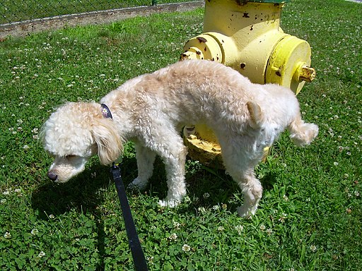
Communicating with Urine
Why do dogs pee on fire hydrants? Besides “having to go,” they are marking their territory with chemicals in their urine called pheromones. It’s a form of communication, in which they are “saying” with odors that the yard is theirs and other dogs should stay away. In addition to fire hydrants, dogs may urinate on fence posts, trees, car tires, and many other objects. Urination in dogs, as in people, is usually a voluntary process controlled by the brain. The process of forming urine — which occurs in the kidneys — occurs constantly, and is not under voluntary control. What happens to all the urine that forms in the kidneys? It passes from the kidneys through the other organs of the urinary system, starting with the ureters.
Ureters
As shown in Figure 16.5.2, ureters are tube-like structures that connect the kidneys with the urinary bladder. They are paired structures, with one ureter for each kidney. In adults, ureters are between 25 and 30 cm (about 10–12 in) long and about 3 to 4 mm in diameter.
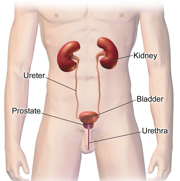
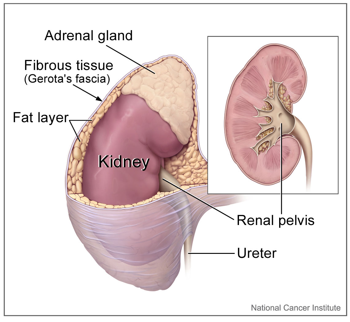
Each ureter arises in the pelvis of a kidney (the renal pelvis in Figure 16.5.3). It then passes down the side of the kidney, and finally enters the back of the bladder. At the entrance to the bladder, the ureters have sphincters that prevent the backflow of urine.
The walls of the ureters are composed of multiple layers of different types of tissues. The innermost layer is a special type of epithelium, called transitional epithelium. Unlike the epithelium lining most organs, transitional epithelium is capable of stretching and does not produce mucus. It lines much of the urinary system, including the renal pelvis, bladder, and much of the urethra, in addition to the ureters. Transitional epithelium allows these organs to stretch and expand as they fill with urine or allow urine to pass through. The next layer of the ureter walls is made up of loose connective tissue containing elastic fibres, nerves, and blood and lymphatic vessels. After this layer are two layers of smooth muscles, an inner circular layer, and an outer longitudinal layer. The smooth muscle layers can contract in waves of peristalsis to propel urine down the ureters from the kidneys to the urinary bladder. The outermost layer of the ureter walls consists of fibrous tissue.
Urinary Bladder
The urinary bladder is a hollow, muscular, and stretchy organ that rests on the pelvic floor. It collects and stores urine from the kidneys before the urine is eliminated through urination. As shown in Figure 16.5.4, urine enters the urinary bladder from the ureters through two ureteral openings on either side of the back wall of the bladder. Urine leaves the bladder through a sphincter called the internal urethral sphincter. When the sphincter relaxes and opens, it allows urine to flow out of the bladder and into the urethra.
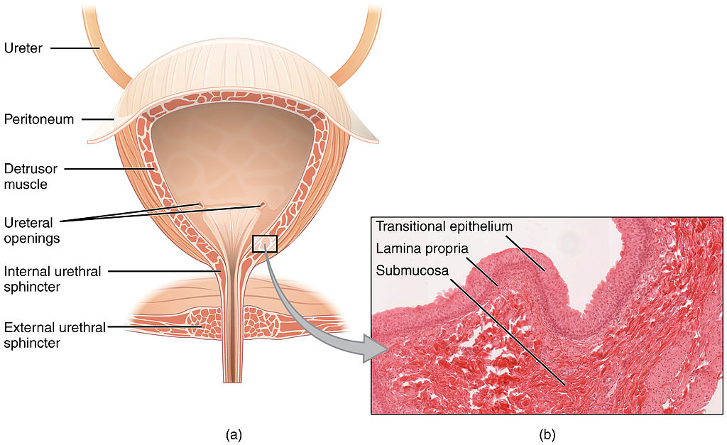
Like the ureters, the bladder is lined with transitional epithelium, which can flatten out and stretch as needed as the bladder fills with urine. The next layer (lamina propria) is a layer of loose connective tissue, nerves, and blood and lymphatic vessels. This is followed by a submucosa layer, which connects the lining of the bladder with the detrusor muscle in the walls of the bladder. The outer covering of the bladder is peritoneum, which is a smooth layer of epithelial cells that lines the abdominal cavity and covers most abdominal organs.
The detrusor muscle in the wall of the bladder is made of smooth muscle fibres controlled by both the autonomic and somatic nervous systems. As the bladder fills, the detrusor muscle automatically relaxes to allow it to hold more urine. When the bladder is about half full, the stretching of the walls triggers the sensation of needing to urinate. When the individual is ready to void, conscious nervous signals cause the detrusor muscle to contract, and the internal urethral sphincter to relax and open. As a result, urine is forcefully expelled out of the bladder and into the urethra.
Urethra
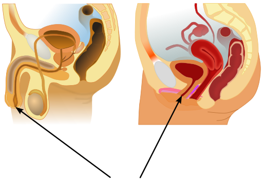
The urethra is a tube that connects the urinary bladder to the external urethral orifice, which is the opening of the urethra on the surface of the body. As shown in Figure 16.5.5, the urethra in males travels through the penis, so it is much longer than the urethra in females. In males, the urethra averages about 20 cm (about 7.8 in) long, whereas in females, it averages only about 4.8 cm (about 1.9 in) long. In males, the urethra carries semen (as well as urine), but in females, it carries only urine. In addition, in males, the urethra passes through the prostate gland (part of the reproductive system) which is absent in women.
Like the ureters and bladder, the proximal (closer to the bladder) two-thirds of the urethra are lined with transitional epithelium. The distal (farther from the bladder) third of the urethra is lined with mucus-secreting epithelium. The mucus helps protect the epithelium from urine, which is corrosive. Below the epithelium is loose connective tissue, and below that are layers of smooth muscle that are continuous with the muscle layers of the urinary bladder. When the bladder contracts to forcefully expel urine, the smooth muscle of the urethra relaxes to allow the urine to pass through.
In order for urine to leave the body through the external urethral orifice, the external urethral sphincter must relax and open. This sphincter is a striated muscle that is controlled by the somatic nervous system, so it is under conscious, voluntary control in most people (exceptions are infants, some elderly people, and patients with certain injuries or disorders). The muscle can be held in a contracted state and hold in the urine until the person is ready to urinate. Following urination, the smooth muscle lining the urethra automatically contracts to re-establish muscle tone, and the individual consciously contracts the external urethral sphincter to close the external urethral opening.
16.5 Summary
- Ureters are tube-like structures that connect the kidneys with the urinary bladder. Each ureter arises at the renal pelvis of a kidney and travels down through the abdomen to the urinary bladder. The walls of the ureter contain smooth muscle that can contract to push urine through the ureter by peristalsis. The walls are lined with transitional epithelium that can expand and stretch.
- The urinary bladder is a hollow, muscular organ that rests on the pelvic floor. It is also lined with transitional epithelium. The function of the bladder is to collect and store urine from the kidneys before the urine is eliminated through urination. Filling of the bladder triggers the sensation of needing to urinate. When a conscious decision to urinate is made, the detrusor muscle in the bladder wall contracts and forces urine out of the bladder and into the urethra.
- The urethra is a tube that connects the urinary bladder to the external urethral orifice. Somatic nerves control the sphincter at the distal end of the urethra. This allows the opening of the sphincter for urination to be under voluntary control.
16.5 Review Questions
- What are ureters? Describe the location of the ureters relative to other urinary tract organs.
- Identify layers in the walls of a ureter. How do they contribute to the ureter’s function?
- Describe the urinary bladder. What is the function of the urinary bladder?
-
- How does the nervous system control the urinary bladder?
- What is the urethra?
- How does the nervous system control urination?
- Identify the sphincters that are located along the pathway from the ureters to the external urethral orifice.
- What are two differences between the male and female urethra?
- When the bladder muscle contracts, the smooth muscle in the walls of the urethra _________ .
16.5 Explore More
The taboo secret to better health | Molly Winter, TED. 2016.
What Happens When You Hold Your Pee? SciShow, 2016.
Attributions
Figure 16.5.1
Cliche by Jackie on Wikimedia Common s is used under a CC BY 2.0 (https://creativecommons.org/licenses/by/2.0) license.
Figure 16.5.2
Urinary System Male by BruceBlaus on Wikimedia Commons is used under a CC BY-SA 4.0 (https://creativecommons.org/licenses/by-sa/4.0) license.
Figure 16.5.3
Adrenal glands on Kidney by NCI Public Domain by Alan Hoofring (Illustrator) /National Cancer Institute (photo ID 4355) on Wikimedia Commons is in the public domain (https://en.wikipedia.org/wiki/Public_domain).
Figure 16.5.4
2605_The_Bladder by OpenStax College on Wikimedia Commons is used under a CC BY 3.0 (https://creativecommons.org/licenses/by/3.0) license. (Micrograph originally provided by the Regents of the University of Michigan Medical School © 2012.)
Figure 16.5.5
512px-Male_and_female_urethral_openings.svg by andrybak (derivative work) on Wikimedia Commons is used under a CC BY-SA 3.0 (https://creativecommons.org/licenses/by-sa/3.0) license. (Original: Male anatomy blank.svg: alt.sex FAQ, derivative work: Tsaitgaist Female anatomy with g-spot.svg: Tsaitgaist.)
References
Betts, J. G., Young, K.A., Wise, J.A., Johnson, E., Poe, B., Kruse, D.H., Korol, O., Johnson, J.E., Womble, M., DeSaix, P. (2013, June 19). Figure 25.4 Bladder (a) Anterior cross section of the bladder. (b) The detrusor muscle of the bladder (source: monkey tissue) LM × 448 [digital image]. In Anatomy and Physiology (Section 7.3). OpenStax. https://openstax.org/books/anatomy-and-physiology/pages/25-2-gross-anatomy-of-urine-transport
SciShow. (2016, January 22). What happens when you hold your pee? YouTube. https://www.youtube.com/watch?v=dg4_deyHLvQ&feature=youtu.be
TED. (2016, September 2). The taboo secret to better health | Molly Winter. YouTube. https://www.youtube.com/watch?v=2Brajdazp1o&feature=youtu.be
A secreted or excreted chemical factor that triggers a social response in members of the same species. Pheromones are chemicals capable of acting like hormones outside the body of the secreting individual, to impact the behavior of the receiving individuals.
Actions which take place according to the one's desire or are under control.
The central nervous system organ inside the skull that is the control center of the nervous system.
A muscular, tube-like organ of the urinary system that moves urine by peristalsis from a kidney to the bladder.
A distinctive pattern of smooth muscle contractions that propels foodstuffs distally through the esophagus and intestines.
A sac-like organ that stores urine until it is excreted from the body.
A liquid waste product of the body that is formed by the kidneys and excreted by the other organs of the urinary system.
One of a pair of organs of the excretory and urinary systems that filters wastes and excess water out of blood and forms urine.
The process in which urine leaves the body through the external urethral orifice.
division of the peripheral nervous system that controls involuntary activities
A division of the peripheral nervous system that controls voluntary activities.
A tube-like organ of the urinary system that carries urine out of the body from the bladder and, in males, also carries semen out of the body.
The funnel-like end of a ureter where it enters the kidney and where urine collects before it is transported through the ureter.
An involuntary, nonstriated muscle that is found in the walls of internal organs such as the stomach.
A ring of muscles that can contract to close off an opening between structures, such as between the esophagus and stomach.
