10.6 Nails
Created by CK-12 Foundation/Adapted by Christine Miller

Nail Art
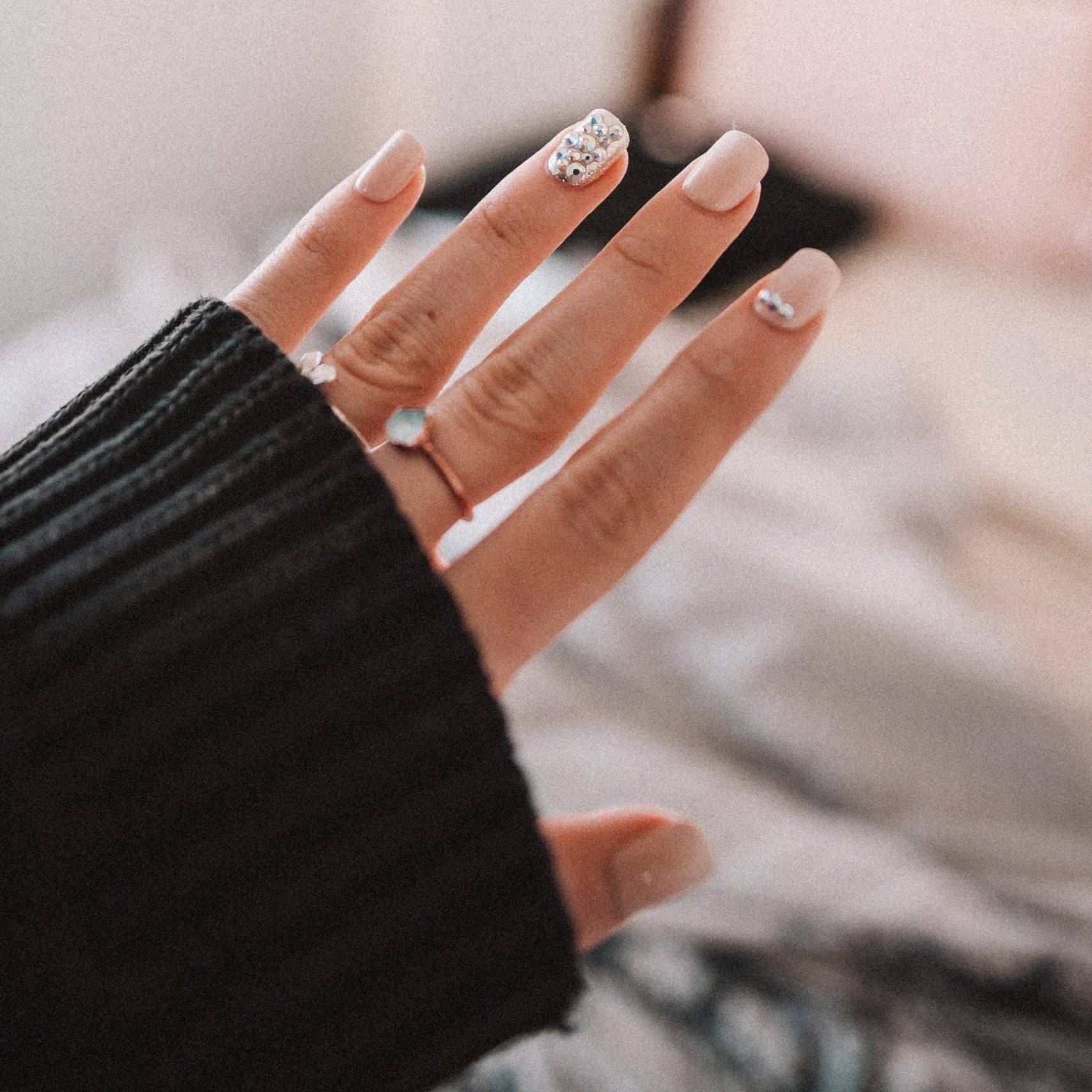
Painting nails with coloured polish for aesthetic reasons is nothing new. In fact, there is evidence of this practice dating back to at least 3000 BCE. Today, painting and otherwise decorating the nails is big business, with annual revenues in the billions of dollars in North America alone! With all the attention (and money) given to nails as decorative objects, it’s easy to forget that they also have important biological functions.
What Are Nails?
Nails are accessory organs of the skin. They are made of sheets of dead keratinocytes and are found on the far (or distal) ends of the fingers and toes. The keratin in nails makes them hard, but flexible. Nails serve a number of purposes, including protecting the digits, enhancing sensations, and acting as tools.
Nail Anatomy

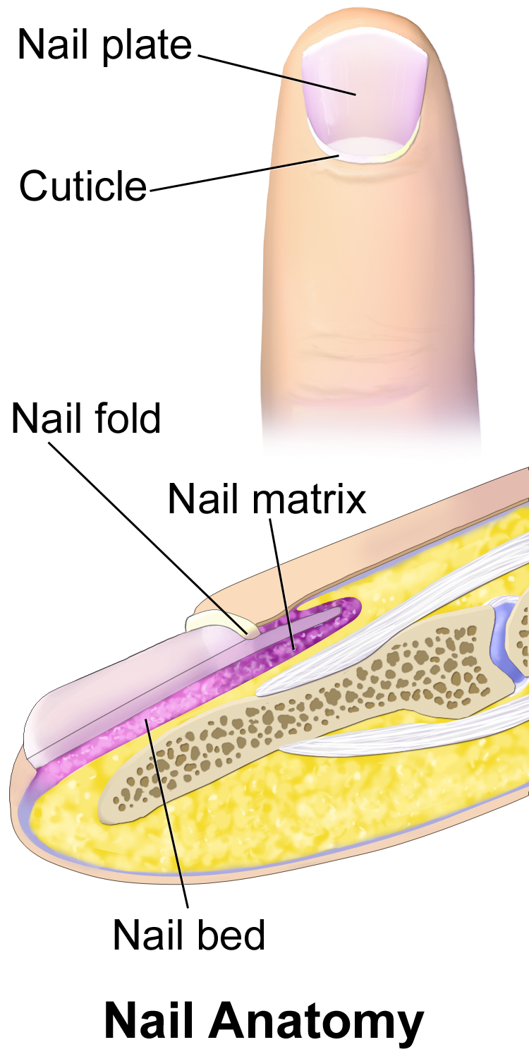
A nail has three main parts: the root, plate, and free margin. Other structures around or under the nail include the nail bed, cuticle, and nail fold. These structures are shown in Figure 10.6.2.
- The nail root is the portion of the nail found under the surface of the skin at the near (or proximal) end of the nail. It is where the nail begins.
- The nail plate (or body) is the portion of the nail that is external to the skin. It is the visible part of the nail.
- The free margin is the portion of the nail that protrudes beyond the distal end of the finger or toe. This is the part that is cut or filed to keep the nail trimmed.
- The nail bed is the area of skin under the nail plate. It is pink in colour, due to the presence of capillaries in the dermis.
- The cuticle is a layer of dead epithelial cells that overlaps and covers the edge of the nail plate. It helps to seal the edges of the nail to prevent infection of the underlying tissues.
- The nail fold is a groove in the skin in which the side edges of the nail plate are embedded.
Nail Growth
Nails grow from a deep layer of living epidermal tissue, known as the nail matrix, at the proximal end of the nail (see the bottom of the diagram in Figure 10.6.2). The nail matrix surrounds the nail root. It contains stem cells that divide to form keratinocytes, which are cells that produce keratin and make up the nail.
Formation of the Nail Root and Nail Plate
The keratinocytes produced by the nail matrix accumulate to form tough, hard, translucent sheets of dead cells filled with keratin. The sheets make up the nail root, which slowly grows out of the skin and becomes the nail plate when it reaches the skin surface. As the nail grows longer, the cells of the nail root and nail plate are pushed toward the distal end of the finger or toe by new cells being formed in the nail matrix. The upper epidermal cells of the nail bed also move along with the nail plate as it grows toward the tip of the digit.
The proximal end of the nail plate near the root has a whitish crescent shape called the lunula. This is where a small amount of the nail matrix is visible through the nail plate. The lunula is most pronounced in the nails of the thumbs, and may not be visible in the nails of the little fingers.
Rate of Nail Growth
Nails grow at an average rate of 3 mm a month. Fingernails, however, grow up to four times as fast as toenails. If a fingernail is lost, it takes between three and six months to regrow completely, whereas a toenail takes between 12 and 18 months to regrow. The actual rate of growth of an individual’s nails depends on many factors, including age, sex, season, diet, exercise level, and genes. If protected from breaking, nails can sometimes grow to be very long. The Chinese doctor in the photo below (Figure 10.6.3) has very long nails on two fingers of his left hand. This picture was taken in 1920 in China, where having long nails was a sign of aristocracy since it implied that one was wealthy enough to not have to do physical labour.

Functions of Nails
Both fingernails and toenails protect the soft tissues of the fingers and toes from injury. Fingernails also serve to enhance sensation and precise movements of the fingertips through the counter-pressure exerted on the pulp of the fingers by the nails. In addition, fingernails can function as several different types of tools. For example, they enable a fine precision grip like tweezers, and can also be used for cutting and scraping.
Nails and Health
Healthcare providers, particularly EMTs, often examine the fingernail beds as a quick and easy indicator of oxygen saturation of the blood, or the amount of blood reaching the extremities. If the nail beds are bluish or purple, it is generally a sign of low oxygen saturation. To see if blood flow to the extremities is adequate, a blanch test may be done. In this test, a fingernail is briefly depressed to turn the nail bed white by forcing the blood out of its capillaries. When the pressure is released, the pink colour of the nail bed should return within a second or two if there is normal blood flow. If the return to a pink colour is delayed, then it can be an indicator of low blood volume, due to dehydration or shock.

How the visible portion of the nails appears can be used as an indicator of recent health status. In fact, nails have been used as diagnostic tools for hundreds — if not thousands — of years. Nail abnormalities, such as deep grooves, brittleness, discolouration, or unusually thin or thick nails, may indicate various illnesses, nutrient deficiencies, drug reactions, or other health problems.
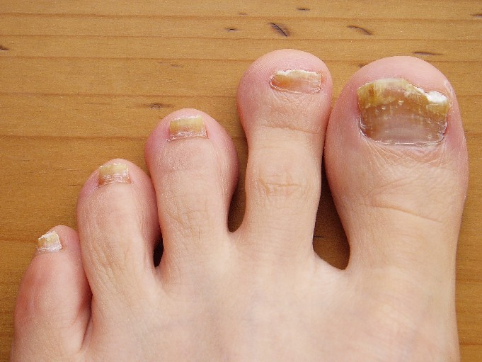
Nails — especially toenails — are common sites of fungal infections (shown in Figure 10.6.4), causing nails to become thickened and yellowish in colour. Toenails are more often infected than fingernails because they are often confined in shoes, which creates a dark, warm, moist environment where fungi can thrive. Toes also tend to have less blood flow than fingers, making it harder for the immune system to detect and stop infections in toenails.
Although nails are harder and tougher than skin, they are also more permeable. Harmful substances may be absorbed through the nails and cause health problems. Some of the substances that can pass through the nails include the herbicide Paraquat, fungicidal agents such as miconazole (e.g., Monistat), and sodium hypochlorite, which is an ingredient in common household bleach. Care should be taken to protect the nails from such substances when handling or immersing the hands in them by wearing latex or rubber gloves.
Feature: Reliable Sources

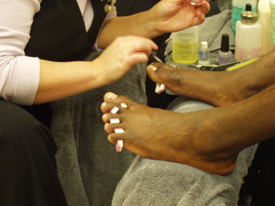
Do you get regular manicures or pedicures from a nail technician? If so, there is a chance that you are putting your health at risk. Nail tools that are not properly disinfected between clients may transmit infections from one person to another. Cutting the cuticles with scissors may create breaks in the skin that let infective agents enter the body. Products such as acrylics, adhesives, and UV gels that are applied to the nails may be harmful, especially if they penetrate the nails and enter the skin.
Use the Internet to find several reliable sources that address the health risks of professional manicures or pedicures. Try to find answers to the following questions:
- What training and certification are required for professional nail technicians?
- What licenses and inspections are required for nail salons?
- What hygienic practices should be followed in nail salons to reduce the risk of infections being transmitted to clients?
- Which professional nail products are potentially harmful to the human body and which are safer?
- How likely is it to have an adverse health consequence when you get a professional manicure or pedicure?
- What steps can you take to ensure that a professional manicure or pedicure is safe?
10.6 Summary
- Nails are accessory organs of the skin, consisting of sheets of dead, keratin-filled keratinocytes. The keratin in nails makes them hard, but flexible.
- A nail has three main parts: the nail root (which is under the epidermis), the nail plate (which is the visible part of the nail), and the free margin (which is the distal edge of the nail). Other structures under or around a nail include the nail bed, cuticle, and nail fold.
- A nail grows from a deep layer of living epidermal tissues — called the nail matrix — at the proximal end of the nail. Stem cells in the nail matrix keep dividing to allow nail growth, forming first the nail root and then the nail plate as the nail continues to grow longer and emerges from the epidermis.
- Fingernails grow faster than toenails. Actual rates of growth depend on many factors, such as age, sex, and season.
- Functions of nails include protecting the digits, enhancing sensations and precise movements of the fingertips, and acting as tools.
- The colour of the nail bed can be used to quickly assess oxygen and blood flow in a patient. How the nail plate grows out can reflect recent health problems, such as illness or nutrient deficiency.
- Nails — and especially toenails — are prone to fungus infections. Nails are more permeable than skin and can absorb several harmful substances, such as herbicides.
10.6 Review Questions
- What are nails?
-
- Explain why most of the nail plate looks pink.
- Describe a lunula.
- Explain how a nail grows.
- Identify three functions of nails.
- Give several examples of how nails are related to health.
- What is the cuticle of the nail composed of? What is the function of the cuticle? Why is it a bad idea to cut the cuticle during a manicure?
- Is the nail plate composed of living or dead cells?
10.6 Explore More
Longest Fingernails – Guinness World Records 60th Anniversary,
Guinness World Records, 2014.
5 Things Your Nails Can Say About Your Health, SciShow, 2015.
Claws vs. Nails – Matthew Borths, TED-Ed, 2019.
Attributions
Figure 10.6.1
Nails by allison-christine-vPrqHSLdF28 [photo] by allison christine on Unsplash is used under the Unsplash License (https://unsplash.com/license).
Figure 10.6.2
Blausen_0406_FingerNailAnatomy by BruceBlaus on Wikimedia Commons is used under a CC BY 3.0 (https://creativecommons.org/licenses/by/3.0) license.
Figure 10.6.3
Chinese_doctor_with_long_finger_nails_(an_aristocrat),_ca.1920_(CHS-249) by Pierce, C.C. (Charles C.), 1861-1946 from the USC Digital Library on Wikimedia Commons is in the public domain (https://en.wikipedia.org/wiki/public_domain).
Figure 10.6.4
Toenail fungus Nagelpilz-3 by Pepsyrock on Wikimedia Commons is released into the public domain (https://en.wikipedia.org/wiki/public_domain).
Figure 10.6.5
OLYMPUS DIGITAL CAMERA by Stoive at the English language Wikipedia, on Wikimedia Commons is used under a CC BY-SA 3.0 (http://creativecommons.org/licenses/by-sa/3.0/) license.
References
Blausen.com staff. (2014). Medical gallery of Blausen Medical 2014. WikiJournal of Medicine 1 (2). DOI:10.15347/wjm/2014.010. ISSN 2002-4436.
Guiness World Records. (2014, December 8). Longest fingernails – Guinness World Records 60th Anniversary. YouTube. https://www.youtube.com/watch?v=G35kPhbUZdg
SciShow. (2015, September 14). 5 things your nails can say about your health. YouTube. https://www.youtube.com/watch?v=aTSVHwzkYI4
TED-Ed. (2019, October 29). Claws vs. nails – Matthew Borths. YouTube. https://www.youtube.com/watch?v=7w2gCBL1MCg
accessory organ of the skin made of sheets of dead keratinocytes at the distal ends of the fingers and toes
The major organ of the integumentary system that covers and protects the body and helps maintain homeostasis, for example, by regulating body temperature.
A type of epithelial cell found in the skin, hair, and nails that produces keratin.
A portion of a nail that is under the surface of the skin at the proximal end of the nail.
visible part of a nail that is external to the skin
The part of the nail that protrudes beyond the end of the finger or toe; the part that is cut or filed to keep the nail trimmed.
The pink skin under the nail plate visible through the nail.
A layer of clear skin located along the bottom edge of your finger or toe. The cuticle function is to protect new nails from bacteria when they grow out from the nail root. The area around the cuticle is delicate. It can get dry, damaged, and infected.
The tissue that encloses the nail matrix at the root of the nail.
A deep layer of epidermal tissue at the proximal end of a nail where nail growth occurs.
A tough, fibrous protein in skin, hair, and nails.
The white area at the base of a fingernail.

