12.7 Case Study Conclusion: Needing to Relax
Created by CK-12 Foundation/Adapted by Christine Miller

Case Study Conclusion: Needing to Relax
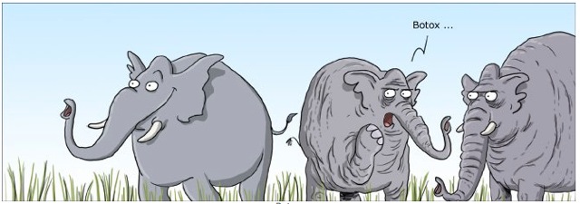
As you learned in the beginning of this chapter, botulinum toxin — one form of which is sold under the brand name Botox — does much more than smooth out wrinkles. It can be used to treat a number of disorders involving excessive muscle contraction, including cervical dystonia. You also learned that cervical dystonia, which Edward suffers from, causes abnormal, involuntary muscle contractions of the neck. This results in jerky movements of the head and neck, and/or a sustained abnormal tilt to the head. It is often painful and can significantly interfere with a person’s life.

How could a toxin actually help treat a muscular disorder? The botulinum toxin is produced by the soil bacterium, Clostridium botulinum, and it is the cause of the potentially deadly disease called botulism. Botulism is often a foodborne illness, commonly caused by foods that are improperly canned. Other forms of botulism are caused by wound infections, or occur when infants consume spores of the bacteria from soil or honey.
Botulism can be life-threatening, because it paralyzes muscles throughout the body, including those involved in breathing. When a very small amount of botulinum toxin is injected carefully into specific muscles by a trained medical professional, however, it can be useful in inhibiting unwanted muscle contractions.
For cosmetic purposes, botulinum toxin injected into the facial muscles relaxes them to reduce the appearance of wrinkles. When used to treat cervical dystonia, it is injected into the muscles of the neck to inhibit excessive muscle contractions. For many patients, this helps relieve the abnormal positioning, movements, and pain associated with the disorder. The effect is temporary, so the injections must be repeated every three to four months to keep the symptoms under control.
How does botulinum toxin inhibit muscle contraction? First, recall how skeletal muscle contraction works. A motor neuron instructs skeletal muscle fibres to contract at a synapse between them called the neuromuscular junction. A nerve impulse called an action potential travels down to the axon terminal of the motor neuron, where it causes the release of the neurotransmitter acetylcholine (ACh) from synaptic vesicles. The ACh travels across the synaptic cleft and binds to ACh receptors on the muscle fibre, signaling the muscle fibre to contract. According to the sliding filament theory, the contraction of the muscle fibre occurs due to the sliding of myosin and actin filaments across each other. This causes the Z discs of the sacromeres to move closer together, shortening the sacromeres and causing the muscle fibre to contract.
If you wanted to inhibit muscle contraction, at what points could you theoretically interfere with this process? Inhibiting the action potential in the motor neuron, the release of ACh, the activity of ACh receptors, or the sliding filament process in the muscle fibre would all theoretically impair this process and inhibit muscle contraction. For example, in the disease myasthenia gravis, the function of the ACh receptors is impaired, causing a lack of sufficient muscle contraction. As you have learned, this results in muscle weakness that can eventually become life-threatening. Botulinum toxin works by inhibiting the release of ACh from the motor neurons, thereby removing the signal instructing the muscles to contract.
Fortunately, Edward’s excessive muscle contractions and associated pain improved significantly thanks to botulinum toxin injections. Although cervical dystonia cannot currently be cured, botulinum toxin injections have improved the quality of life for many patients with this and other disorders involving excessive involuntary muscle contractions.
As you have learned in this chapter, our muscular system allows us to do things like make voluntary movements, digest our food, and pump blood through our bodies. Whether they are in your arm, heart, stomach, or blood vessels, muscle tissue works by contracting. But as you have seen here, too much contraction can be a very bad thing. Fortunately, scientists and physicians have found a way to put a potentially deadly toxin — and wrinkle-reducing treatment — to excellent use as a medical treatment for some muscular system disorders.
Chapter 12 Summary
In this chapter, you learned about the muscular system. Specifically, you learned that:
- The muscular system consists of all the muscles of the body. There are three types of muscle: skeletal muscle (which is attached to bones by tendons and enables voluntary body movements), cardiac muscle (which makes up the walls of the heart and makes it beat) and smooth muscle (which is found in the walls of internal organs and other internal structures and controls their movements).
- Muscles are organs composed mainly of muscle cells, which may also be called muscle fibres or myocytes. Muscle cells are specialized for the function of contracting, which occurs when protein filaments inside the cells slide over one another using energy from ATP. Muscle tissue is the only type of tissue that has cells with the ability to contract.
- Muscles can grow larger, or hypertrophy. This generally occurs through increased use, although hormonal or other influences can also play a role. Muscles can also grow smaller, or atrophy. This may occur through lack of use, starvation, certain diseases, or aging. In both hypertrophy and atrophy, the size — but not the number — of muscle fibres changes. The size of muscles is the main determinant of muscle strength.
- Skeletal muscles need the stimulus of motor neurons to contract, and to move the body, they need the skeletal system to act upon.
- Skeletal muscle is the most common type of muscle tissue in the human body. To move bones in opposite directions, skeletal muscles often consist of pairs of muscles that work in opposition to one another to move bones in different directions at joints.
- Skeletal muscle fibres are bundled together in units called muscle fascicles, which are bundled together to form individual skeletal muscles. Skeletal muscles also have connective tissue supporting and protecting the muscle tissue.
-
- Each skeletal muscle fibre consists of a bundle of myofibrils, which are bundles of protein filaments. The filaments are arranged in repeating units called sarcomeres, which are the basic functional units of skeletal muscles. Skeletal muscle tissue is striated, because of the pattern of sarcomeres in its fibres.
- Skeletal muscle fibres can be divided into two types, called slow-twitch and fast-twitch fibres. Slow-twitch fibres are used mainly in aerobic endurance activities (such as long-distance running). Fast-twitch fibres are used mainly for non-aerobic, strenuous activities (such as sprinting). Proportions of the two types of fibres vary from muscle to muscle and person to person.
- Smooth muscle tissue is found in the walls of internal organs and vessels. When smooth muscles contract, they help the organs and vessels carry out their functions. The pattern of smooth muscle contraction to move substances through body tubes is called peristalsis. Contractions of smooth muscles are involuntary and controlled by the autonomic nervous system, hormones, and other substances.
-
- Cells of smooth muscle tissue are not striated because they lack sarcomeres, but the cells contract in the same basic way as striated muscle cells. Unlike striated muscle, smooth muscle can sustain very long-term contractions and maintain its contractile function, even when stretched.
- Cardiac muscle tissue is found only in the wall of the heart. When cardiac muscle contracts, the heart beats and pumps blood. Contractions of cardiac muscle are involuntary, like those of smooth muscles. They are controlled by electrical impulses from specialized cardiac cells.
-
- Like skeletal muscle, cardiac muscle is striated because its filaments are arranged in sarcomeres. The exact arrangement, however, differs, making cardiac and skeletal muscle tissues look different from one another.
- The heart is the muscle that performs the greatest amount of physical work in the course of a lifetime. Its cells contain a great many mitochondria to produce ATP for energy and to help the heart resist fatigue.
- A muscle contraction is an increase in the tension or a decrease in the length of a muscle. A muscle contraction is isometric if muscle tension changes, but muscle length remains the same. It is isotonic if muscle length changes, but muscle tension remains the same.
-
- A skeletal muscle contraction begins with electrochemical stimulation of a muscle fibre by a motor neuron. This occurs at a chemical synapse called a neuromuscular junction. The neurotransmitter acetylcholine diffuses across the synaptic cleft and binds to receptors on the muscle fibre. This initiates a muscle contraction.
- Once stimulated, the protein filaments within the skeletal muscle fibre slide past each other to produce a contraction. The sliding filament theory is the most widely accepted explanation for how this occurs. According to this theory, thick myosin filaments repeatedly attach to and pull on thin actin filaments, thus shortening sarcomeres.
- Crossbridge cycling is a cycle of molecular events that underlies the sliding filament theory. Using energy in ATP, myosin heads repeatedly bind with and pull on actin filaments. This moves the actin filaments toward the center of a sarcomere, shortening the sarcomere and causing a muscle contraction.
- The ATP needed for a muscle contraction comes first from ATP already available in the cell, and more is generated from creatine phosphate. These sources are quickly used up. Glucose and glycogen can be broken down to form ATP and pyruvate. Pyruvate can then be used to produce ATP in aerobic respiration if oxygen is available, or it can be used in anaerobic respiration if oxygen is not available.
- Physical exercise is defined as any bodily activity that enhances or maintains physical fitness and overall health. Activities such as household chores may even count as physical exercise! Current recommendations for adults are 30 minutes of moderate exercise a day.
- Aerobic exercise is any physical activity that uses muscles at less than their maximum contraction strength, but for long periods of time. This type of exercise uses a relatively high percentage of slow-twitch muscle fibres that consume large amounts of oxygen. Aerobic exercises increase cardiovascular endurance, and include cycling and brisk walking.
- Anaerobic exercise is any physical activity that uses muscles at close to their maximum contraction strength, but for short periods of time. This type of exercise uses a relatively high percentage of fast-twitch muscle fibres that consume small amounts of oxygen. Anaerobic exercises increase muscle and bone mass and strength, and they include push-ups and sprinting.
- Flexibility exercise is any physical activity that stretches and lengthens muscles, thereby improving range of motion and reducing risk of injury. Examples include stretching and yoga.
- Many studies have shown that physical exercise is positively correlated with a diversity of physical, mental, and emotional health benefits. Physical exercise also increases quality of life and life expectancy.
-
- Many of the benefits of exercise may come about because contracting muscles release hormones called myokines, which promote tissue repair and growth and have anti-inflammatory effects.
- Physical exercise can reduce risk factors for cardiovascular disease, including hypertension and excess body weight. Physical exercise can also increase factors associated with cardiovascular health, such as mechanical efficiency of the heart.
- Physical exercise has been shown to offer protection from dementia and other cognitive problems, perhaps because it increases blood flow or neurotransmitters in the brain, among other potential effects.
- Numerous studies suggest that regular aerobic exercise works as well as pharmaceutical antidepressants in treating mild-to-moderate depression, possibly because it increases synthesis of natural euphoriants in the brain.
- Research shows that physical exercise generally improves sleep for most people, and helps sleep disorders, such as insomnia. Other health benefits of physical exercise include better immune system function and reduced risk of type 2 diabetes and obesity.
- There is great variation in individual responses to exercise, partly due to genetic differences in proportions of slow-twitch and fast-twitch muscle fibres. People with more slow-twitch fibres may be able to develop greater endurance from aerobic exercise, whereas people with more fast-twitch fibres may be able to develop greater muscle size and strength from anaerobic exercise.
- Some adverse effects may occur if exercise is extremely intense and the body is not given proper rest between exercise sessions. Many people who overwork their muscles develop delayed onset muscle soreness (DOMS), which may be caused by tiny tears in muscle fibres.
- Musculoskeletal disorders are injuries that occur in muscles or associated tissues (such as tendons) because of biomechanical stresses. The disorders may be caused by sudden exertion, over-exertion, repetitive motions, and similar stresses.
-
- A muscle strain is an injury in which muscle fibres tear as a result of overstretching. First aid for a muscle strain includes the five steps represented by the acronym PRICE (protection, rest, ice, compression, and elevation). Medications for inflammation and pain (such as NSAIDs) may also be used.
- Tendinitis is inflammation of a tendon that occurs when it is over-extended or worked too hard without rest. Tendinitis may also be treated with PRICE and NSAIDs.
- Carpal tunnel syndrome is a biomechanical problem that occurs in the wrist when the median nerve becomes compressed between carpal bones. It may occur with repetitive use, a tumor, or trauma to the wrist. It may cause pain, numbness, and eventually — if untreated — muscle wasting in the thumb and first two fingers of the hand.
- Neuromuscular disorders are systemic disorders that occur because of problems with the nervous control of muscle contractions, or with the muscle cells themselves.
-
- Muscular dystrophy is a genetic disorder caused by defective proteins in muscle cells. It is characterized by progressive skeletal muscle weakness and death of muscle tissues.
- Myasthenia gravis is a genetic neuromuscular disorder characterized by fluctuating muscle weakness and fatigue. More muscles are affected, and muscles become increasingly weakened as the disorder progresses. Myasthenia gravis most often occurs because immune system antibodies block acetylcholine receptors on muscle cells, and because of the actual loss of acetylcholine receptors.
- Parkinson’s disease is a degenerative disorder of the central nervous system that mainly affects the muscular system and movement. It occurs because of the death of neurons in the midbrain. Characteristic signs of the disorder are muscle tremor, muscle rigidity, slowness of movement, and postural instability. Dementia and depression also often characterize advanced stages of the disease.
As you saw in this chapter, muscles need oxygen to provide enough ATP for most of their activities. In fact, all of the body’s systems require oxygen, and also need to remove waste products, such as carbon dioxide. In the next chapter, you will learn about how the respiratory system obtains and distributes oxygen throughout the body, as well as how it removes wastes, such as carbon dioxide.
Chapter 12 Review
-
- What are tendons? Name a muscular system disorder involving tendons
- Describe the relationship between muscles, muscle fibres, and fascicles.

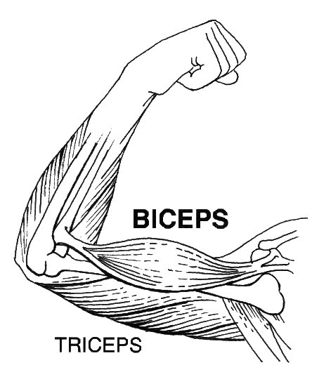
- The biceps and triceps muscles are shown above. Answer the following questions about these arm muscles.
- When the biceps contract and become shorter (as in the picture above), what kind of motion does this produce in the arm?
- Is the situation described in part (a) more likely to be an isometric or isotonic contraction? Explain your answer.
- If the triceps were to then contract, which way would the arm move?
- What are Z discs? What happens to them during muscle contraction?
- What is the function of mitochondria in muscle cells? Which type of muscle fibre has more mitochondria — slow-twitch or fast-twitch?
- What is the difference between primary and secondary Parkinson’s disease?
- Why can carpal tunnel syndrome cause muscle weakness in the hands?
Attributions
Figure 12.7.1
Botox, he whispered by Michael Reuter on Flickr is used under a CC BY 2.0 (https://creativecommons.org/licenses/by/2.0/) license.
Figure 12.7.2
botulism by jason wilson on Flickr is used under a CC BY 2.0 (https://creativecommons.org/licenses/by/2.0/) license.
Reference
Pearson Scott Foresman. (2020, April 14). File:Biceps (PSF).jpg [digital image]. Wikimedia Commons. https://commons.wikimedia.org/w/index.php?title=File:Biceps_(PSF).jpg&oldid=411251538. [Public Domain (https://en.wikipedia.org/wiki/Public_domain)]
A drug prepared from the bacterial toxin botulin, used medically to treat certain muscular conditions and cosmetically to remove wrinkles by temporarily paralyzing facial muscles.
The body system responsible for the movement of the human body. Attached to the bones of the skeletal system are about 700 named muscles that make up roughly half of a person's body weight. Each of these muscles is a discrete organ constructed of skeletal muscle tissue, blood vessels, tendons, and nerves.
Voluntary, striated muscle that is attached to bones of the skeleton and helps the body move.
Involuntary, striated muscle found only in the walls of the heart; also called myocardium.
An involuntary, nonstriated muscle that is found in the walls of internal organs such as the stomach.
A long, thin muscle cell that has the ability to contract.
A type of muscle cell that makes up smooth muscle tissue.
A complex organic chemical that provides energy to drive many processes in living cells, e.g. muscle contraction, nerve impulse propagation, and chemical synthesis. Found in all forms of life, ATP is often referred to as the "molecular unit of currency" of intracellular energy transfer.
An increase in the size of a structure, such as an increase in the size of a muscle through exercise.
The decrease in the size of a structure, such as a decrease in the size of a muscle through non-use.
A type of neuron that carries nerve impulses from the central nervous system to muscles and glands; also called efferent neuron.
The body system composed of bones and cartilage and performs the following critical functions for the human body: supports the body. The skeletal system facilitates movement, protects internal organs, and produces blood cells.
A structure where two or more bones of the skeleton come together.
Long filaments that run parallel to each other to form muscle (myo) fibers. The muscle fibers are single multinucleated cells that combine to form the muscle. Myofibrils are made up of repeating subunits called sarcomeres.
The basic functional unit of skeletal and cardiac muscles, containing actin and myosin protein filaments that slide over one another to produce a shortening of the sarcomere resulting in a muscle contraction.
A type of skeletal muscle cell that is mainly responsible for aerobic activities such as long-distance running.
A type of skeletal muscle cell that is mainly responsible for anaerobic activities such as weight lifting.
Relating to, involving, or requiring free oxygen.
A distinctive pattern of smooth muscle contractions that propels foodstuffs distally through the esophagus and intestines.
Actions which are not under one's conscious control.
division of the peripheral nervous system that controls involuntary activities
A hormone is a signaling molecule produced by glands in multicellular organisms that target distant organs to regulate physiology and behavior.
A muscular organ in the chest that pumps blood through blood vessels when it contracts.
A double-membrane-bound organelle found in most eukaryotic organisms. Mitochondria convert oxygen and nutrients into adenosine triphosphate (ATP). ATP is the chemical energy "currency" of the cell that powers the cell's metabolic activities.
Referring to a muscle contraction in which muscle tension increases but muscle length remains the same.
Referring to a muscle contraction in which muscle length decreases but muscle tension remains the same.
A theory that explains muscle contraction by the sliding of myosin filaments over actin filaments within muscle fibers.
A fibrous protein that forms (together with actin) the contractile filaments of muscle cells and is also involved in motion in other types of cells.
A protein that forms (together with myosin) the contractile filaments of muscle cells, and is also involved in motion in other types of cells.
A sequence of molecular events that forms crossbridges between myosin and actin filaments in muscle fibers, allowing for muscle contraction. "Heads" on the myosin filaments essentially form a connection with specific locations on the actin, and then the head bends in order to pull the myosin strand along the actin to shorten the sarcomere.
An organic compound of creatine and phosphate, also known as phosphocreatine, which when hydrolyzed (split apart) releases energy for muscle contraction.
Glucose (also called dextrose) is a simple sugar with the molecular formula C6H12O6. Glucose is the most abundant monosaccharide, a subcategory of carbohydrates. Glucose is mainly made by plants and most algae during photosynthesis from water and carbon dioxide, using energy from sunlight.
A multi-branched polysaccharide of glucose that serves as a form of energy storage in animals, fungi, and bacteria.
The process of producing cellular energy involving oxygen. Cells break down food in the mitochondria in a long, multi-step process that produces roughly 36 ATP. The first step in is glycolysis, the second is the Krebs cycle and the third is the electron transport system.
Respiration using electron acceptors other than molecular oxygen. Although oxygen is not the final electron acceptor, the process still uses a respiratory electron transport chain.
Any physical activity in which muscles are used well below their maximum contraction strength but for a relatively long period of time, consuming a large amount of oxygen.
Any physical activity in which muscles are used at close to their maximum contraction strength but for a relatively short period to time, consuming a small amount of oxygen.
Any physical activity that stretches and lengthens muscles.
One of several hundred cytokines or other small proteins produced and released by muscle cells (myocytes) in response to muscular contractions with an endocrine function.
A persistently high blood pressure, generally defined as 140/90 mm Hg or higher.
Abnormal or excessive fat accumulation that presents a risk to health. Obesity has been more precisely defined by the National Institutes of Health (the NIH) as a BMI (Body Mass Index) of 30 and above.
A chronic or persistent disorder of the mental processes caused by brain disease or injury and marked by memory disorders, personality changes, and impaired reasoning.
A common and serious medical illness that negatively affects how you feel, the way you think and how you act. Fortunately, it is also treatable. Depression causes feelings of sadness and/or a loss of interest in activities once enjoyed. It can lead to a variety of emotional and physical problems and can decrease a person’s ability to function at work and at home.
A type of drug that tends to induce a feeling or state of intense excitement and happiness.
An injury to muscles or tendons caused by biomechanical stresses.
An injury in which muscle fibers tear due to overstretching of a muscle.
Inflammation of a tendon when it is over-extended or worked too hard without rest.
A musculoskeletal disorder that occurs when a nerve becomes compressed between carpal bones in the wrist, leading to reduced innervation of the thumb and first two fingers.
A disorder that occurs due to problems with the nervous control of muscle contractions or with muscle cells themselves.
A genetic neuromuscular disorder caused by defective proteins in muscle cells and characterized by death of skeletal muscles and progressive weakness.
A genetic neuromuscular disorder caused by the immune system blocking or destroying acetylcholine receptors on muscle cells and characterized by progressive muscle weakness and fatigue.
A degenerative brain disorder caused by progressive death of neurons in the midbrain, resulting in muscular symptoms of tremor, rigidity, slowness of movement, and postural instability.

